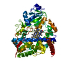
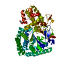
BIOMOLECULE: 1 THIS ENTRY CONTAINS THE CRYSTALLOGRAPHIC ASYMMETRIC UNIT WHICH CONSISTS OF 2 CHAIN(S) ...BIOMOLECULE: 1 THIS ENTRY CONTAINS THE CRYSTALLOGRAPHIC ASYMMETRIC UNIT WHICH CONSISTS OF 2 CHAIN(S). SEE REMARK 350 FOR INFORMATION ON GENERATING THE BIOLOGICAL MOLECULE(S). SIZE EXCLUSION CHROMATOGRAPHY WITH STATIC LIGHT SCATTERING SUPPORTS A MIXTURE OF DIMER AND TETRAMER IN SOLUTION. THE PISA SERVER ALSO PREDICTS BOTH THE TETRAMER AND DIMER TO BE STABLE. THE TETRAMER IS DESCRIBED IN REMARK 350.
Remark 999
SEQUENCE: (1) THE CONSTRUCT WAS EXPRESSED WITH A PURIFICATION TAG MGSDKIHHHHHHENLYFQG. THE TAG WAS ...SEQUENCE: (1) THE CONSTRUCT WAS EXPRESSED WITH A PURIFICATION TAG MGSDKIHHHHHHENLYFQG. THE TAG WAS REMOVED WITH TEV PROTEASE LEAVING ONLY A GLYCINE (0) FOLLOWED BY THE TARGET SEQUENCE. (2) THE SEQUENCE OF THE PROTEIN WAS NOT AVAILABLE AT THE UNP DATABASE AT THE TIME OF PROCESSING. (3) THE SEQUENCE IS AVAILABLE FROM GENBANK UNDER ACCESSION ID ZP_00243239.1 AND FROM THE UNIPROT ARCHIVE UNDER ACCESSION ID UPI00003CCEF6.
SIZE EXCLUSION CHROMATOGRAPHY WITH STATIC LIGHT SCATTERING SUPPORTS A MIXTURE OF DIMER AND TETRAMER IN SOLUTION. THE PISA SERVER ALSO PREDICTS BOTH THE TETRAMER AND DIMER TO BE STABLE.
Mass: 15856.126 Da / Num. of mol.: 2 Source method: isolated from a genetically manipulated source Source: (gene. exp.) Methylibium petroleiphilum (bacteria) / Strain: PM1 Description: Methylobium petroleophilum is another scientific name of the source organism Gene: ZP_00243239.1 / Production host: Escherichia coli (E. coli)
Mass: 18.015 Da / Num. of mol.: 202 / Source method: isolated from a natural source / Formula: H2O
-
Details
Has protein modification
Y
-
Experimental details
-
Experiment
Experiment
Method: X-RAY DIFFRACTION / Number of used crystals: 1
-
Sample preparation
Crystal
Density Matthews: 2.03 Å3/Da / Density % sol: 39.33 % Description: DATA FROM TWO CRYSTALS WERE USED FOR THE STRUCTURE DETERMINATION. ONE CRYSTAL WAS USED FOR MAD PHASING EXPERIMENTS AND TRACING AT A RESOLUTION OF 2.0 ANGTROMS. THE 2.0 ANGSTROM MODEL WAS ...Description: DATA FROM TWO CRYSTALS WERE USED FOR THE STRUCTURE DETERMINATION. ONE CRYSTAL WAS USED FOR MAD PHASING EXPERIMENTS AND TRACING AT A RESOLUTION OF 2.0 ANGTROMS. THE 2.0 ANGSTROM MODEL WAS REFINED TO AN ENHANCED RESOLUTION OF 1.50 ANGSTROMS USING DATA FROM A SEPARATE CRYSTAL WITH THE 2 ANGSTROM MAD PHASES FROM THE FIRST CRYSTAL USED AS PHASE RESTRAINTS.
Resolution: 1.5→29.437 Å / Num. obs: 42659 / % possible obs: 99.7 % / Biso Wilson estimate: 16.91 Å2 / Rmerge(I) obs: 0.094 / Net I/σ(I): 9.49
Reflection shell
Resolution (Å)
Rmerge(I) obs
Mean I/σ(I) obs
Num. measured obs
Diffraction-ID
% possible all
1.5-1.55
0.732
1.9
25145
1,2
97.7
1.55-1.62
0.639
2.3
32670
1,2
100
1.62-1.69
0.566
2.7
27786
1,2
100
1.69-1.78
0.439
3.4
29288
1,2
100
1.78-1.89
0.313
4.4
27933
1,2
99.9
1.89-2.04
0.218
5.9
28091
1,2
99.8
2.04-2.24
0.207
9.2
52079
1,2
99.9
2.24-2.56
0.142
12.4
54678
1,2
99.9
2.56-3.23
0.083
19
55655
1,2
99.8
3.23-29.4
0.057
33.6
57993
1,2
99.5
-
Phasing
Phasing
Method: MAD
-
Processing
Software
Name
Version
Classification
NB
MolProbity
3beta29
modelbuilding
SHELX
phasing
REFMAC
5.2.0019
refinement
XSCALE
datascaling
PDB_EXTRACT
2
dataextraction
XDS
datareduction
SHELXD
phasing
autoSHARP
phasing
Refinement
Method to determine structure: MAD / Resolution: 1.5→29.437 Å / Cor.coef. Fo:Fc: 0.963 / Cor.coef. Fo:Fc free: 0.951 / SU B: 2.939 / SU ML: 0.058 / TLS residual ADP flag: LIKELY RESIDUAL / Cross valid method: THROUGHOUT / σ(F): 0 / ESU R: 0.08 / ESU R Free: 0.08 Stereochemistry target values: MAXIMUM LIKELIHOOD WITH PHASES Details: (1). HYDROGENS HAVE BEEN ADDED IN RIDING POSITIONS. (2). ATOM RECORD CONTAINS RESIDUAL B FACTORS ONLY. (3). A MET-INHIBITION PROTOCOL WAS USED FOR SELENOMETHIONINE INCORPORATION DURING ...Details: (1). HYDROGENS HAVE BEEN ADDED IN RIDING POSITIONS. (2). ATOM RECORD CONTAINS RESIDUAL B FACTORS ONLY. (3). A MET-INHIBITION PROTOCOL WAS USED FOR SELENOMETHIONINE INCORPORATION DURING PROTEIN EXPRESSION. THE OCCUPANCY OF THE SE ATOMS IN THE MSE RESIDUES WAS REDUCED TO 0.75 TO ACCOUNT FOR THE REDUCED SCATTERING POWER DUE TO PARTIAL S-MET INCORPORATION. (4). A ZN ATOM ON EACH OF THE TWO SUBUNITS IN THE ASYMMETRIC UNIT IS COORDINATED TO THE SIDE CHAIN OF HIS 59, HIS 101, AND ACETATE. A ZINC ATOM ON SUBUNIT B IS COORDINATED TO THE SIDE CHAINS OF GLU 136, ASP 137 AND FOUR WATER MOLECULES. ANOMALOUS DIFFERENCE FOURIERS AND X-RAY FLUORESCENCE EXPERIMENTS SUPPORT THE ASSIGNMENT OF THE ZINC IONS. (5). UNEXPLAINED ELECTRON DENSITIES OBSERVED NEAR RESIDUE 111 ON THE A SUBUNIT AND RESIDUES 141-143 ON THE B SUBUNIT WERE NOT MODELED. (6). FOUR MOLECULES OF POLYETHYLENE GLYCOL 200 (PG4) USED AS A CRYOPROTECTANT, FIVE ACETATE (ACT) AND ONE CL ION FROM THE CRYSTALLIZATION BUFFER WERE MODELED INTO THE STRUCTURE.
Rfactor
Num. reflection
% reflection
Selection details
Rfree
0.21
2177
5.1 %
RANDOM
Rwork
0.181
-
-
-
obs
0.183
42575
99.76 %
-
Solvent computation
Ion probe radii: 0.8 Å / Shrinkage radii: 0.8 Å / VDW probe radii: 1.2 Å / Solvent model: MASK
Displacement parameters
Biso mean: 16.549 Å2
Baniso -1
Baniso -2
Baniso -3
1-
0.28 Å2
0 Å2
0 Å2
2-
-
0.28 Å2
0 Å2
3-
-
-
-0.56 Å2
Refinement step
Cycle: LAST / Resolution: 1.5→29.437 Å
Protein
Nucleic acid
Ligand
Solvent
Total
Num. atoms
2170
0
58
202
2430
Refine LS restraints
Refine-ID
Type
Dev ideal
Dev ideal target
Number
X-RAY DIFFRACTION
r_bond_refined_d
0.017
0.022
2373
X-RAY DIFFRACTION
r_bond_other_d
0.001
0.02
1577
X-RAY DIFFRACTION
r_angle_refined_deg
1.706
1.949
3219
X-RAY DIFFRACTION
r_angle_other_deg
0.935
3
3843
X-RAY DIFFRACTION
r_dihedral_angle_1_deg
7
5
304
X-RAY DIFFRACTION
r_dihedral_angle_2_deg
36.395
23.846
91
X-RAY DIFFRACTION
r_dihedral_angle_3_deg
12.429
15
324
X-RAY DIFFRACTION
r_dihedral_angle_4_deg
14.698
15
5
X-RAY DIFFRACTION
r_chiral_restr
0.106
0.2
320
X-RAY DIFFRACTION
r_gen_planes_refined
0.009
0.02
2724
X-RAY DIFFRACTION
r_gen_planes_other
0.001
0.02
499
X-RAY DIFFRACTION
r_nbd_refined
0.233
0.2
498
X-RAY DIFFRACTION
r_nbd_other
0.202
0.2
1517
X-RAY DIFFRACTION
r_nbtor_refined
0.189
0.2
1142
X-RAY DIFFRACTION
r_nbtor_other
0.091
0.2
1260
X-RAY DIFFRACTION
r_xyhbond_nbd_refined
0.229
0.2
171
X-RAY DIFFRACTION
r_metal_ion_refined
0.215
0.2
9
X-RAY DIFFRACTION
r_symmetry_vdw_refined
0.162
0.2
29
X-RAY DIFFRACTION
r_symmetry_vdw_other
0.248
0.2
115
X-RAY DIFFRACTION
r_symmetry_hbond_refined
0.215
0.2
30
X-RAY DIFFRACTION
r_mcbond_it
2.194
3
1549
X-RAY DIFFRACTION
r_mcbond_other
0.628
3
606
X-RAY DIFFRACTION
r_mcangle_it
2.784
5
2382
X-RAY DIFFRACTION
r_scbond_it
4.311
8
997
X-RAY DIFFRACTION
r_scangle_it
5.221
11
837
LS refinement shell
Resolution: 1.5→1.539 Å / Total num. of bins used: 20
In the structure databanks used in Yorodumi, some data are registered as the other names, "COVID-19 virus" and "2019-nCoV". Here are the details of the virus and the list of structure data.
Jan 31, 2019. EMDB accession codes are about to change! (news from PDBe EMDB page)
EMDB accession codes are about to change! (news from PDBe EMDB page)
The allocation of 4 digits for EMDB accession codes will soon come to an end. Whilst these codes will remain in use, new EMDB accession codes will include an additional digit and will expand incrementally as the available range of codes is exhausted. The current 4-digit format prefixed with “EMD-” (i.e. EMD-XXXX) will advance to a 5-digit format (i.e. EMD-XXXXX), and so on. It is currently estimated that the 4-digit codes will be depleted around Spring 2019, at which point the 5-digit format will come into force.
The EM Navigator/Yorodumi systems omit the EMD- prefix.
Related info.:Q: What is EMD? / ID/Accession-code notation in Yorodumi/EM Navigator
Yorodumi is a browser for structure data from EMDB, PDB, SASBDB, etc.
This page is also the successor to EM Navigator detail page, and also detail information page/front-end page for Omokage search.
The word "yorodu" (or yorozu) is an old Japanese word meaning "ten thousand". "mi" (miru) is to see.
Related info.:EMDB / PDB / SASBDB / Comparison of 3 databanks / Yorodumi Search / Aug 31, 2016. New EM Navigator & Yorodumi / Yorodumi Papers / Jmol/JSmol / Function and homology information / Changes in new EM Navigator and Yorodumi
 Movie
Movie Controller

 Yorodumi
 Open data
 Basic information
 Components
 Keywords
 Function and homology information
 Methylibium petroleiphilum (bacteria)
 X-RAY DIFFRACTION /
  Authors
 Citation
 Structure visualization
 Downloads & links
 Other downloads
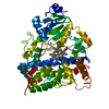


 PDBj
PDBj Assembly
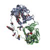


 Mass: 65.409 Da / Num. of mol.: 3 / Source method: obtained synthetically / Formula: Zn
 Mass: 35.453 Da / Num. of mol.: 1 / Source method: obtained synthetically / Formula: Cl
 Mass: 59.044 Da / Num. of mol.: 5 / Source method: obtained synthetically / Formula: C2H3O2
 Mass: 194.226 Da / Num. of mol.: 4 / Source method: obtained synthetically / Formula: C8H18O5 / Comment: precipitant*YM
 Sample preparation
 Processing