- PDB-2ns1: Crystal structure of the e. coli ammonia channel AMTB complexed w... -
+
Open data
ID or keywords:
Loading...
-
Basic information
Entry
Database: PDB / ID: 2ns1
Title
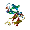
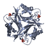
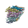
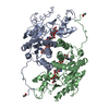
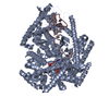
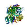
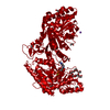
Crystal structure of the e. coli ammonia channel AMTB complexed with the signal transduction protein GLNK
Components
Ammonia channel
Nitrogen regulatory protein P-II 2
Keywords
TRANSPORT PROTEIN/SIGNALING PROTEIN / PROTEIN-PROTEIN COMPLEX / MEMBRANE PROTEIN / AMMONIA / CHANNEL / REGULATORY / INHIBITOR / SIGNAL PROTEIN / ADP / BOG / Structural Genomics / PSI-2 / Protein Structure Initiative / Center for Structures of Membrane Proteins / CSMP / TRANSPORT PROTEIN-SIGNALING PROTEIN COMPLEX
Function / homology
Function and homology information
positive regulation of nitrogen utilization / ammonium transmembrane transport / ammonium channel activity / regulation of nitrogen utilization / enzyme regulator activity / carbon dioxide transport / ATP binding / identical protein binding / plasma membrane / cytosol Similarity search - Function
Ammonium transporter, conserved site / Ammonium transporters signature. / Nitrogen regulatory protein P-II, urydylation site / P-II protein uridylation site. / Ammonium transporter / Ammonium transporter fold / Ammonium transporter AmtB like domains / Ammonium transporter AmtB-like domain / Ammonium Transporter Family / Nitrogen regulatory protein PII, conserved site ...Ammonium transporter, conserved site / Ammonium transporters signature. / Nitrogen regulatory protein P-II, urydylation site / P-II protein uridylation site. / Ammonium transporter / Ammonium transporter fold / Ammonium transporter AmtB like domains / Ammonium transporter AmtB-like domain / Ammonium Transporter Family / Nitrogen regulatory protein PII, conserved site / P-II protein C-terminal region signature. / Nitrogen regulatory protein P-II / P-II protein family profile. / Ammonium/urea transporter / Nitrogen regulatory protein PII / Nitrogen regulatory protein P-II / Alpha-Beta Plaits - #120 / Nitrogen regulatory PII-like, alpha/beta / Nitrogen regulatory protein PII/ATP phosphoribosyltransferase, C-terminal / Alpha-Beta Plaits / 2-Layer Sandwich / Orthogonal Bundle / Mainly Alpha / Alpha Beta Similarity search - Domain/homology
In the structure databanks used in Yorodumi, some data are registered as the other names, "COVID-19 virus" and "2019-nCoV". Here are the details of the virus and the list of structure data.
Jan 31, 2019. EMDB accession codes are about to change! (news from PDBe EMDB page)
EMDB accession codes are about to change! (news from PDBe EMDB page)
The allocation of 4 digits for EMDB accession codes will soon come to an end. Whilst these codes will remain in use, new EMDB accession codes will include an additional digit and will expand incrementally as the available range of codes is exhausted. The current 4-digit format prefixed with “EMD-” (i.e. EMD-XXXX) will advance to a 5-digit format (i.e. EMD-XXXXX), and so on. It is currently estimated that the 4-digit codes will be depleted around Spring 2019, at which point the 5-digit format will come into force.
The EM Navigator/Yorodumi systems omit the EMD- prefix.
Related info.:Q: What is EMD? / ID/Accession-code notation in Yorodumi/EM Navigator
Yorodumi is a browser for structure data from EMDB, PDB, SASBDB, etc.
This page is also the successor to EM Navigator detail page, and also detail information page/front-end page for Omokage search.
The word "yorodu" (or yorozu) is an old Japanese word meaning "ten thousand". "mi" (miru) is to see.
Related info.:EMDB / PDB / SASBDB / Comparison of 3 databanks / Yorodumi Search / Aug 31, 2016. New EM Navigator & Yorodumi / Yorodumi Papers / Jmol/JSmol / Function and homology information / Changes in new EM Navigator and Yorodumi
 Movie
Movie Controller

 Yorodumi
 Open data
 Basic information
 Components
 Keywords
 Function and homology information
 X-RAY DIFFRACTION /
  Authors
 Citation
 Structure visualization
 Downloads & links
 Other downloads
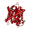


 PDBj
PDBj Assembly


 Type: D-saccharide / Mass: 292.369 Da / Num. of mol.: 8


 Mass: 122.143 Da / Num. of mol.: 2 / Source method: obtained synthetically / Formula: C4H12NO3 / Comment: pH buffer*YM
 Mass: 427.201 Da / Num. of mol.: 1 / Source method: obtained synthetically / Formula: C10H15N5O10P2 / Comment: ADP, energy-carrying molecule*YM
 Sample preparation
 / Beamline: 8.3.1 / Wavelength: 1.1159 Å
 Processing