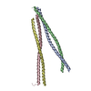
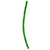
登録情報 データベース : PDB / ID : 2no2タイトル Crystal structure of the DLLRKN-containing coiled-coil domain of Huntingtin-interacting protein 1 Huntingtin-interacting protein 1 キーワード / / / / 機能・相同性 分子機能 ドメイン・相同性 構成要素
/ / / / / / / / / / / / / / / / / / / / / / / / / / / / / / / / / / / / / / / / / / / / / / / / / / / / / / / / / / / / / / / / / / / / / / / / / / / / / / / / / 生物種 Homo sapiens (ヒト)手法 / / / 解像度 : 2.8 Å データ登録者 Ybe, J.A. / Mishra, S. / Helms, S. / Nix, J. ジャーナル : J.Mol.Biol. / 年 : 2007タイトル : Crystal structure at 2.8 A of the DLLRKN-containing coiled-coil domain of huntingtin-interacting protein 1 (HIP1) reveals a surface suitable for clathrin light chain binding著者 : Ybe, J.A. / Mishra, S. / Helms, S. / Nix, J. 履歴 登録 2006年10月24日 登録サイト / 処理サイト 改定 1.0 2007年10月30日 Provider / タイプ 改定 1.1 2011年7月13日 Group 改定 1.2 2023年12月27日 Group / Database referencesカテゴリ chem_comp_atom / chem_comp_bond ... chem_comp_atom / chem_comp_bond / database_2 / struct_ref_seq_dif Item / _database_2.pdbx_database_accession / _struct_ref_seq_dif.details
すべて表示 表示を減らす
 ムービー
ムービー コントローラー


 データを開く
 基本情報
基本情報 要素
要素 キーワード
キーワード 機能・相同性情報
 Homo sapiens (ヒト)
 X線回折 /
  データ登録者
 引用
引用 構造の表示
構造の表示 ダウンロードとリンク
 その他のダウンロード
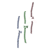
 PDBj
PDBj

 集合体
集合体

 分子量: 18.015 Da / 分子数: 14 / 由来タイプ: 天然 / 式: H2O
 試料調製
試料調製 / ビームライン: 4.2.2 / 波長: 0.97878, 0.97863, 0.96409
 解析
解析