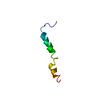
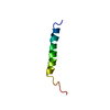
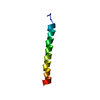
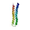
- PDB-2n7i: NMR structure of the prolactin receptor transmembrane domain -
+
Open data
ID or keywords:
Loading...
-
Basic information
Entry
Database: PDB / ID: 2n7i
Title
NMR structure of the prolactin receptor transmembrane domain
Components
Prolactin receptor
Keywords
HORMONE RECEPTOR / transmembrane domain / membrane protein / receptor
Function / homology
Function and homology information
prolactin receptor activity / activation of transmembrane receptor protein tyrosine kinase activity / regulation of epithelial cell differentiation / steroid biosynthetic process / activation of Janus kinase activity / mammary gland epithelial cell differentiation / prostate gland growth / cellular response to granulocyte macrophage colony-stimulating factor stimulus / Prolactin receptor signaling / cytokine binding ...prolactin receptor activity / activation of transmembrane receptor protein tyrosine kinase activity / regulation of epithelial cell differentiation / steroid biosynthetic process / activation of Janus kinase activity / mammary gland epithelial cell differentiation / prostate gland growth / cellular response to granulocyte macrophage colony-stimulating factor stimulus / Prolactin receptor signaling / cytokine binding / peptide hormone binding / mammary gland alveolus development / Growth hormone receptor signaling / regulation of cell adhesion / cell surface receptor signaling pathway via JAK-STAT / positive regulation of B cell proliferation / lactation / embryo implantation / endosome lumen / response to bacterium / cytokine-mediated signaling pathway / positive regulation of cold-induced thermogenesis / signaling receptor complex / external side of plasma membrane / positive regulation of cell population proliferation / lipid binding / protein kinase binding / negative regulation of apoptotic process / cell surface / extracellular region / metal ion binding / plasma membrane Similarity search - Function
Long hematopoietin receptor, single chain, conserved site / Growth hormone/erythropoietin receptor, ligand binding / Erythropoietin receptor, ligand binding / Long hematopoietin receptor, single chain family signature. / : / Fibronectin type 3 domain / Fibronectin type-III domain profile. / Fibronectin type III / Fibronectin type III superfamily / Immunoglobulin-like fold Similarity search - Domain/homology
Mass: 3960.745 Da / Num. of mol.: 1 / Fragment: Helical transmembrane domain residues 230-264 Source method: isolated from a genetically manipulated source Source: (gene. exp.) Homo sapiens (human) / Gene: PRLR / Production host: Escherichia coli (E. coli) / Strain (production host): BL21(DE3) / References: UniProt: P16471
-
Experimental details
-
Experiment
Experiment
Method: SOLUTION NMR
NMR experiment
Conditions-ID
Experiment-ID
Solution-ID
Type
1
1
2
2D 1H-15N HSQC
1
2
3
3D HNHA
1
3
3
2D 1H-15N HSQC
1
4
1
2D 1H-15N HSQC
1
5
2
2D 1H-15N HSQC
1
6
1
3D HNCO
1
7
1
3D HN(CA)CB
1
8
1
3DCBCA(CO)NH
1
9
2
3D 1H-15N NOESY
1
10
2
2D 1H-13C HSQC aliphatic
1
11
2
2D 1H-13C HSQC aromatic
-
Sample preparation
Details
Solution-ID
Contents
Solvent system
1
0.8 mM [U-13C; U-15N] PRLRTMD, 2 mM DSS, 0.05 % sodium azide, 560 mM DHPC, 20 mM sodium phosphate, 4 mM TCEP, 50 mM sodium chloride, 90% H2O/10% D2O
90% H2O/10% D2O
2
1 mM [U-13C; U-15N] PRLRTMD, 2 mM DSS, 0.05 % sodium azide, 700 mM [U-2H] DHPC, 20 mM sodium phosphate, 4 mM TCEP, 50 mM sodium chloride, 90% H2O/10% D2O
90% H2O/10% D2O
3
0.7 mM [U-15N] PRLRTMD, 2 mM DSS, 0.05 % sodium azide, 490 mM DHPC, 20 mM sodium phosphate, 4 mM TCEP, 50 mM sodium chloride, 90% H2O/10% D2O
In the structure databanks used in Yorodumi, some data are registered as the other names, "COVID-19 virus" and "2019-nCoV". Here are the details of the virus and the list of structure data.
Jan 31, 2019. EMDB accession codes are about to change! (news from PDBe EMDB page)
EMDB accession codes are about to change! (news from PDBe EMDB page)
The allocation of 4 digits for EMDB accession codes will soon come to an end. Whilst these codes will remain in use, new EMDB accession codes will include an additional digit and will expand incrementally as the available range of codes is exhausted. The current 4-digit format prefixed with “EMD-” (i.e. EMD-XXXX) will advance to a 5-digit format (i.e. EMD-XXXXX), and so on. It is currently estimated that the 4-digit codes will be depleted around Spring 2019, at which point the 5-digit format will come into force.
The EM Navigator/Yorodumi systems omit the EMD- prefix.
Related info.:Q: What is EMD? / ID/Accession-code notation in Yorodumi/EM Navigator
Yorodumi is a browser for structure data from EMDB, PDB, SASBDB, etc.
This page is also the successor to EM Navigator detail page, and also detail information page/front-end page for Omokage search.
The word "yorodu" (or yorozu) is an old Japanese word meaning "ten thousand". "mi" (miru) is to see.
Related info.:EMDB / PDB / SASBDB / Comparison of 3 databanks / Yorodumi Search / Aug 31, 2016. New EM Navigator & Yorodumi / Yorodumi Papers / Jmol/JSmol / Function and homology information / Changes in new EM Navigator and Yorodumi
 Movie
Movie Controller


 Open data
 Basic information
 Components
 Keywords
 Function and homology information
 Homo sapiens (human)
 Authors
 Citation
 Structure visualization
 Downloads & links
 Other downloads
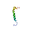

 PDBj
PDBj


 Assembly

 HSQC
HSQC Sample preparation
 Processing