 Movie
Movie Controller

[English] 日本語
 Yorodumi
- PDB-2l56: NMR structure of the GCN4 trigger peptide refined using biased mo... -
+ Open data

- Basic information
| Entry | Database: PDB / ID: 2l56 | ||||||
|---|---|---|---|---|---|---|---|
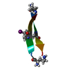
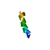
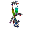
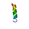
| Title | NMR structure of the GCN4 trigger peptide refined using biased molecular dynamics simulations | ||||||
 Components Components | General control protein GCN4 | ||||||
 Keywords Keywords | TRANSCRIPTION / GCN4 / COILED-COIL / TRIGGER PEPTIDE / MOLECULAR DYNAMICS SIMULATIONS | ||||||
| Method | SOLUTION NMR / molecular dynamics | ||||||
| Model details | occurrence frequencies, model 1 | ||||||
 Authors Authors | Missimer, J.H. / Dolenc, J. / Steinmetz, M.O. / van Gunsteren, W.F. | ||||||
 Citation Citation | Journal: Proc.Natl.Acad.Sci.Usa / Year: 2007 Title: Molecular basis of coiled-coil formation Authors: Steinmetz, M.O. / Jelesarov, I. / Matousek, W.M. / Honnappa, S. / Jahnke, W. / Missimer, J.H. / Frank, S. / Alexandrescu, A.T. / Kammerer, R.A. | ||||||
| History |
| ||||||
| Remark 0 | THIS ENTRY 2L56 REFLECTS AN ALTERNATIVE MODELING OF THE ORIGINAL STRUCTURAL DATA (2OVN.MR) ...THIS ENTRY 2L56 REFLECTS AN ALTERNATIVE MODELING OF THE ORIGINAL STRUCTURAL DATA (2OVN.MR) DETERMINED BY AUTHORS OF THE PDB ENTRY 2OVN: M.O.STEINMETZ,I.JELESAROV,W.M.MATOUSEK,S.HONNAPPA,W.JAHNKE,J.H.MISSIMER,S.FRANK,A.T.ALEXANDRESCU,R.A.KAMMERER. HYDROGEN BOND DISTANCE RESTRAINTS IN 2OVN.MR WERE NOT USED IN THE NEW REFINEMENT PROCEDURE BECAUSE THEY ARE NOT BASED ON THE EXPERIMENTAL NMR DATA. |
- Structure visualization
| Structure viewer | Molecule:  MolmilJmol/JSmol MolmilJmol/JSmol |
|---|
- Downloads & links
-Download
| PDBx/mmCIF format | 2l56.cif.gz | 40.2 KB | Display | PDBx/mmCIF format |
|---|---|---|---|---|
| PDB format | pdb2l56.ent.gz | 31.1 KB | Display | PDB format |
| PDBx/mmJSON format | 2l56.json.gz | Tree view | PDBx/mmJSON format | |
| Others |  Other downloads Other downloads |
-Validation report
| Summary document | 2l56_validation.pdf.gz | 354.1 KB | Display | wwPDB validaton report |
|---|---|---|---|---|
| Full document | 2l56_full_validation.pdf.gz | 407.6 KB | Display | |
| Data in XML | 2l56_validation.xml.gz | 7 KB | Display | |
| Data in CIF | 2l56_validation.cif.gz | 9.3 KB | Display | |
| Arichive directory | https://data.pdbj.org/pub/pdb/validation_reports/l5/2l56ftp://data.pdbj.org/pub/pdb/validation_reports/l5/2l56 | HTTPS FTP |
-Related structure data
| Related structure data | |
|---|---|
| Similar structure data |


-Links
 PDBj
PDBj- Assembly
| Deposited unit | 
| |||||||||
|---|---|---|---|---|---|---|---|---|---|---|
| 1 |
| |||||||||
| NMR ensembles |
|
-Components
| #1: Protein/peptide | Mass: 1911.232 Da / Num. of mol.: 1 / Source method: obtained synthetically / Details: FMOC synthesis |
|---|
-Experimental details
-Experiment
| Experiment | Method: SOLUTION NMR |
|---|
- Processing
| NMR software | Name: GROMOS / Version: 53A6 FORCE FIELD / Developer: van Gunsteren and Berendsen / Classification: refinement |
|---|---|
| Refinement | Method: molecular dynamics / Software ordinal: 1 Details: molecular dynamics simulation with time-averaged NOE distance restraining and local elevation biased 3J-value restraining using 179 NOE distances and 15 3J-coupling constants; the 10 ...Details: molecular dynamics simulation with time-averaged NOE distance restraining and local elevation biased 3J-value restraining using 179 NOE distances and 15 3J-coupling constants; the 10 deposited structures were selected from a set of 20000 trajectory configurations by conformational cluster analysis; the occurrence frequencies of the clusters that correspond to the deposited model structures are the following: cluster 1 32% cluster 2 14% cluster 3 6% cluster 4 6% cluster 5 6% cluster 6 3% cluster 7 3% cluster 8 2% cluster 9 2% cluster 10 2%. Authors state that The deviations of the deposited trajectory structures from the so-called "ideal" values for geometrical and stereochemical quantities are partially due to the biasing force derived from the NOE distance bounds and 3J-coupling constants, and partially due to the finite temperature (278K) of the simulations. The ideal geometric and stereochemical values of the GROMOS force field are close to those of Engh and Huber. |
| NMR representative | Selection criteria: occurrence frequencies |
| NMR ensemble | Conformer selection criteria: conformational cluster analysis Conformers calculated total number: 20000 / Conformers submitted total number: 10 / Representative conformer: 1 |
