 Movie
Movie Controller

[English] 日本語
 Yorodumi
- PDB-2kc8: Structure of E. coli toxin RelE (R81A/R83A) mutant in complex wit... -
+ Open data

- Basic information
| Entry | Database: PDB / ID: 2kc8 | ||||||
|---|---|---|---|---|---|---|---|
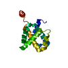
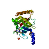
| Title | Structure of E. coli toxin RelE (R81A/R83A) mutant in complex with antitoxin RelBc (K47-L79) peptide | ||||||
 Components Components |
| ||||||
 Keywords Keywords | Toxin/Toxin Repressor / protein-protein complex / toxin RelE / antitoxin RelB / Repressor / Stress response / Toxin / Transcription / Transcription regulation / Toxin-Toxin Repressor COMPLEX | ||||||
| Function / homology |  Function and homology information Function and homology informationtoxin sequestering activity / single-species biofilm formation / regulation of growth / DNA-binding transcription repressor activity / mRNA catabolic process / RNA endonuclease activity / cellular response to amino acid starvation / protein-DNA complex / ribosome binding / endonuclease activity ...toxin sequestering activity / single-species biofilm formation / regulation of growth / DNA-binding transcription repressor activity / mRNA catabolic process / RNA endonuclease activity / cellular response to amino acid starvation / protein-DNA complex / ribosome binding / endonuclease activity / Hydrolases; Acting on ester bonds / negative regulation of translation / transcription cis-regulatory region binding / rRNA binding / response to antibiotic / hydrolase activity / regulation of DNA-templated transcription / DNA-templated transcription Similarity search - Function | ||||||
| Biological species |  | ||||||
| Method | SOLUTION NMR / torsion angle dynamics, simulated annealing | ||||||
 Authors Authors | Li, G. / Zhang, Y. / Inouye, M. / Ikura, M. | ||||||
 Citation Citation | Journal: J.Biol.Chem. / Year: 2009 Title: Inhibitory mechanism of Escherichia coli RelE-RelB toxin-antitoxin module involves a helix displacement near an mRNA interferase active site. Authors: Li, G.Y. / Zhang, Y. / Inouye, M. / Ikura, M. | ||||||
| History |
|
- Structure visualization
| Structure viewer | Molecule: MolmilJmol/JSmol |
|---|
- Downloads & links
-Download
| PDBx/mmCIF format | 2kc8.cif.gz | 832.6 KB | Display | PDBx/mmCIF format |
|---|---|---|---|---|
| PDB format | pdb2kc8.ent.gz | 700.3 KB | Display | PDB format |
| PDBx/mmJSON format | 2kc8.json.gz | Tree view | PDBx/mmJSON format | |
| Others |  Other downloads Other downloads |
-Validation report
| Arichive directory | https://data.pdbj.org/pub/pdb/validation_reports/kc/2kc8ftp://data.pdbj.org/pub/pdb/validation_reports/kc/2kc8 | HTTPS FTP |
|---|
-Related structure data


-Links
 PDBj
PDBj- Assembly
| Deposited unit | 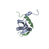
| |||||||||
|---|---|---|---|---|---|---|---|---|---|---|
| 1 |
| |||||||||
| NMR ensembles |
|
-Components
| #1: Protein | Mass: 11356.294 Da / Num. of mol.: 1 / Mutation: R81A, R83A Source method: isolated from a genetically manipulated source Source: (gene. exp.) Description: RelE is expressed with his-tag fusion. The his-tag is removed by thrombin afterward Gene: relE, b1563, JW1555 / Production host: |
|---|---|
| #2: Protein/peptide | Mass: 4135.697 Da / Num. of mol.: 1 / Fragment: UNP residues 47-79 Source method: isolated from a genetically manipulated source Source: (gene. exp.) Description: RelBc (K47-L79) is expressed with GST-tag fusion. The GST-tag is removed by thrombin afterward Gene: relB, b1564, JW1556 / Production host: |
-Experimental details
-Experiment
| Experiment | Method: SOLUTION NMR Details: There is a related deposition of toxin RelE in the peptide free state | ||||||||||||||||||||||||||||||||||||||||||||||||||||||||||||||||||||||||||||||||||||||||||||
|---|---|---|---|---|---|---|---|---|---|---|---|---|---|---|---|---|---|---|---|---|---|---|---|---|---|---|---|---|---|---|---|---|---|---|---|---|---|---|---|---|---|---|---|---|---|---|---|---|---|---|---|---|---|---|---|---|---|---|---|---|---|---|---|---|---|---|---|---|---|---|---|---|---|---|---|---|---|---|---|---|---|---|---|---|---|---|---|---|---|---|---|---|---|
| NMR experiment |
|
 HSQC
HSQC- Sample preparation
| Details |
| ||||||||||||||||||||||||||||||||||||||||||||||||||||||||||||||||||||||||||||||||||||||||||||||||||||
|---|---|---|---|---|---|---|---|---|---|---|---|---|---|---|---|---|---|---|---|---|---|---|---|---|---|---|---|---|---|---|---|---|---|---|---|---|---|---|---|---|---|---|---|---|---|---|---|---|---|---|---|---|---|---|---|---|---|---|---|---|---|---|---|---|---|---|---|---|---|---|---|---|---|---|---|---|---|---|---|---|---|---|---|---|---|---|---|---|---|---|---|---|---|---|---|---|---|---|---|---|---|
| Sample |
| ||||||||||||||||||||||||||||||||||||||||||||||||||||||||||||||||||||||||||||||||||||||||||||||||||||
| Sample conditions | Ionic strength: 0.5 / pH: 6.5 / Pressure: ambient / Temperature: 296.5 K |
-NMR measurement
| NMR spectrometer |
|
|---|
- Processing
| NMR software |
| ||||||||||||||||||||||||||||||||||||||||||||||||||||||||
|---|---|---|---|---|---|---|---|---|---|---|---|---|---|---|---|---|---|---|---|---|---|---|---|---|---|---|---|---|---|---|---|---|---|---|---|---|---|---|---|---|---|---|---|---|---|---|---|---|---|---|---|---|---|---|---|---|---|
| Refinement | Method: torsion angle dynamics, simulated annealing / Software ordinal: 1 | ||||||||||||||||||||||||||||||||||||||||||||||||||||||||
| NMR constraints | NOE constraints total: 3341 / NOE intraresidue total count: 801 / NOE long range total count: 690 / NOE medium range total count: 720 / NOE sequential total count: 782 / Protein chi angle constraints total count: 0 / Protein other angle constraints total count: 0 / Protein phi angle constraints total count: 77 / Protein psi angle constraints total count: 77 | ||||||||||||||||||||||||||||||||||||||||||||||||||||||||
| NMR representative | Selection criteria: lowest energy | ||||||||||||||||||||||||||||||||||||||||||||||||||||||||
| NMR ensemble | Average torsion angle constraint violation: 1.402 ° Conformer selection criteria: structures with the lowest energy Conformers calculated total number: 100 / Conformers submitted total number: 20 / Maximum torsion angle constraint violation: 2.588 ° / Maximum upper distance constraint violation: 0.353 Å / Torsion angle constraint violation method: CNS | ||||||||||||||||||||||||||||||||||||||||||||||||||||||||
| NMR ensemble rms | Distance rms dev: 0.012 Å / Distance rms dev error: 0.001 Å |
