 Movie
Movie Controller

[English] 日本語
 Yorodumi
- PDB-2jx6: Structure and membrane interactions of the antibiotic peptide der... -
+ Open data

- Basic information
| Entry | Database: PDB / ID: 2jx6 | ||||||
|---|---|---|---|---|---|---|---|
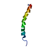
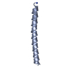
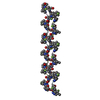
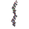
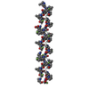
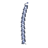
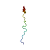
| Title | Structure and membrane interactions of the antibiotic peptide dermadistinctin k by solution and oriented 15N and 31P solid-state NMR spectroscopy | ||||||
 Components Components | Dermadistinctin-K | ||||||
 Keywords Keywords | ANTIMICROBIAL PROTEIN / ALPHA HELIX / AMPHIPATHIC CHARACTER / C-TERMINAL CARBOXYAMIDATION / MEMBRANE PEPTIDE / Amphibian defense peptide / Antibiotic / Antimicrobial / Secreted | ||||||
| Function / homology | Dermaseptin / Dermaseptin / defense response to bacterium / extracellular region / Dermaseptin-DI1 Function and homology information Function and homology information | ||||||
| Biological species | Phyllomedusa distincta (Sao Paulo leaf frog) | ||||||
| Method | SOLUTION NMR / simulated annealing | ||||||
 Authors Authors | Mendonca Moraes, C. / Verly, R.M. / Resende, J.M. / Bemquerer, M.P. / Pilo-Veloso, D. / Valente, A. / Almeida, F.C.L. / Bechinger, B. | ||||||
 Citation Citation | Journal: Biophys.J. / Year: 2009 Title: Structure and membrane interactions of the antibiotic peptide dermadistinctin K by multidimensional solution and oriented 15N and 31P solid-state NMR spectroscopy Authors: Verly, R.M. / de Moraes, C.M. / Resende, J.M. / Aisenbrey, C. / Bemquerer, M.P. / Pilo-Veloso, D. / Valente, A.P. / Almeida, F.C.L. / Bechinger, B. | ||||||
| History |
|
- Structure visualization
| Structure viewer | Molecule: MolmilJmol/JSmol |
|---|
- Downloads & links
-Download
| PDBx/mmCIF format | 2jx6.cif.gz | 183.1 KB | Display | PDBx/mmCIF format |
|---|---|---|---|---|
| PDB format | pdb2jx6.ent.gz | 154.1 KB | Display | PDB format |
| PDBx/mmJSON format | 2jx6.json.gz | Tree view | PDBx/mmJSON format | |
| Others |  Other downloads Other downloads |
-Validation report
| Arichive directory | https://data.pdbj.org/pub/pdb/validation_reports/jx/2jx6ftp://data.pdbj.org/pub/pdb/validation_reports/jx/2jx6 | HTTPS FTP |
|---|
-Related structure data


-Links
 PDBj
PDBj- Assembly
| Deposited unit | 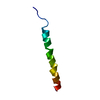
| |||||||||
|---|---|---|---|---|---|---|---|---|---|---|
| 1 |
| |||||||||
| NMR ensembles |
|
-Components
| #1: Protein/peptide | Mass: 3156.699 Da / Num. of mol.: 1 / Source method: obtained synthetically Details: The peptide was synthesized by solid-phase synthesis using fmoc chemistry. The peptide is an antimicrobial peptide that is naturally found from the skin secretion of phyllomedusa distincta, ...Details: The peptide was synthesized by solid-phase synthesis using fmoc chemistry. The peptide is an antimicrobial peptide that is naturally found from the skin secretion of phyllomedusa distincta, a frog species found in brazilian forests. Source: (synth.) Phyllomedusa distincta (Sao Paulo leaf frog) References: UniProt: P83638 |
|---|---|
| Has protein modification | Y |
-Experimental details
-Experiment
| Experiment | Method: SOLUTION NMR | ||||||||||||||||||||
|---|---|---|---|---|---|---|---|---|---|---|---|---|---|---|---|---|---|---|---|---|---|
| NMR experiment |
|
 HSQC
HSQC- Sample preparation
| Details | Contents: 2.0mM dermadistinctin k; trifluoroethanol; H2O 60%, D2O 40% Solvent system: trifluoroethanol; H2O 60%, D2O 40% |
|---|---|
| Sample | Conc.: 2.0 mM / Component: dermadistinctin k |
| Sample conditions | pH: 7 / Pressure: ambient / Temperature: 293.15 K |
-NMR measurement
| NMR spectrometer | Type: Bruker Avance DRX / Manufacturer: Bruker / Model: AVANCE DRX / Field strength: 600 MHz |
|---|
- Processing
| NMR software |
| ||||||||||||||||||||||||||||||||||||||||
|---|---|---|---|---|---|---|---|---|---|---|---|---|---|---|---|---|---|---|---|---|---|---|---|---|---|---|---|---|---|---|---|---|---|---|---|---|---|---|---|---|---|
| Refinement | Method: simulated annealing / Software ordinal: 1 Details: NOE intensities were converted into semi-quantitative distances restrains. The upper limits of the distances thus obtained were 2.8, 3.4 and 5.0A; (for strong, medium, and weak NOEs, ...Details: NOE intensities were converted into semi-quantitative distances restrains. The upper limits of the distances thus obtained were 2.8, 3.4 and 5.0A; (for strong, medium, and weak NOEs, respectively). Structure calculations were performed using the Xplor-NIH software, version 2.17.0 . Starting with the extended structure, 500 structures were generated using a simulated annealing protocol. This was followed by 20000 steps of simulated annealing at 1000 K and a subsequent decrease in tempera ure in 15000 steps in the first slow-cool annealing stage. | ||||||||||||||||||||||||||||||||||||||||
| NMR constraints | NOE constraints total: 294 / NOE intraresidue total count: 178 / NOE long range total count: 0 / NOE medium range total count: 38 / NOE sequential total count: 78 | ||||||||||||||||||||||||||||||||||||||||
| NMR representative | Selection criteria: lowest energy | ||||||||||||||||||||||||||||||||||||||||
| NMR ensemble | Conformer selection criteria: structures with the lowest energy Conformers calculated total number: 500 / Conformers submitted total number: 20 / Representative conformer: 19 |
