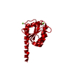
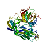
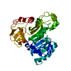
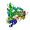
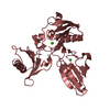
Entry Database : PDB / ID : 2jk1Title Crystal structure of the wild-type HupR receiver domain HYDROGENASE TRANSCRIPTIONAL REGULATORY PROTEIN HUPR1 Keywords / / / / / / / / / / Function / homology Function Domain/homology Component
/ / / / / / / / / / / / / / / / / / / / / / / / / / / / / / / / / / Biological species RHODOBACTER CAPSULATUS (bacteria)Method / / / Resolution : 2.1 Å Authors Davies, K.M. / Lowe, E.D. / Venien-Bryan, C. / Johnson, L.N. Journal : J.Mol.Biol. / Year : 2009Title : The Hupr Receiver Domain Crystal Structure in its Nonphospho and Inhibitory Phospho States.Authors : Davies, K.M. / Lowe, E.D. / Venien-Bryan, C. / Johnson, L.N. History Deposition May 26, 2008 Deposition site / Processing site Revision 1.0 Nov 11, 2008 Provider / Type Revision 1.1 Jul 13, 2011 Group / Version format complianceRevision 1.2 May 8, 2024 Group Data collection / Database references ... Data collection / Database references / Derived calculations / Other Category chem_comp_atom / chem_comp_bond ... chem_comp_atom / chem_comp_bond / database_2 / pdbx_database_status / struct_conn Item _database_2.pdbx_DOI / _database_2.pdbx_database_accession ... _database_2.pdbx_DOI / _database_2.pdbx_database_accession / _pdbx_database_status.status_code_sf / _struct_conn.ptnr1_auth_comp_id / _struct_conn.ptnr1_auth_seq_id / _struct_conn.ptnr1_label_asym_id / _struct_conn.ptnr1_label_atom_id / _struct_conn.ptnr1_label_comp_id / _struct_conn.ptnr1_label_seq_id / _struct_conn.ptnr2_auth_comp_id / _struct_conn.ptnr2_auth_seq_id / _struct_conn.ptnr2_label_asym_id / _struct_conn.ptnr2_label_atom_id / _struct_conn.ptnr2_label_comp_id / _struct_conn.ptnr2_label_seq_id
Show all Show less
 Movie
Movie Controller


 Open data
 Basic information
 Components
 Keywords
 Function and homology information
 RHODOBACTER CAPSULATUS (bacteria)
 X-RAY DIFFRACTION /
  Authors
 Citation
 Structure visualization
 Downloads & links
 Other downloads
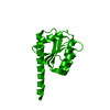

 PDBj
PDBj

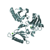
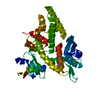
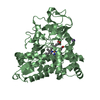
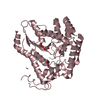
 Assembly
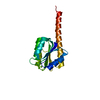


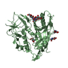
 Mass: 24.305 Da / Num. of mol.: 1 / Source method: obtained synthetically / Formula: Mg
 Mass: 18.015 Da / Num. of mol.: 58 / Source method: isolated from a natural source / Formula: H2O
 Sample preparation
 / Beamline: ID14-2 / Wavelength: 0.933
  Processing