 Movie
Movie Controller

[English] 日本語
 Yorodumi
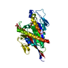
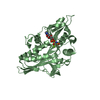
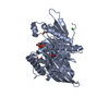
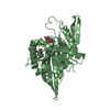
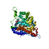
- PDB-2itg: CATALYTIC DOMAIN OF HIV-1 INTEGRASE: ORDERED ACTIVE SITE IN THE F... -
+ Open data

- Basic information
| Entry | Database: PDB / ID: 2itg | ||||||
|---|---|---|---|---|---|---|---|
| Title | CATALYTIC DOMAIN OF HIV-1 INTEGRASE: ORDERED ACTIVE SITE IN THE F185H CONSTRUCT | ||||||
 Components Components | HUMAN IMMUNODEFICIENCY VIRUS-1 INTEGRASE | ||||||
 Keywords Keywords | DNA INTEGRATION / AIDS / POLYPROTEIN / HYDROLASE / ENDONUCLEASE / POLYNUCLEOTIDYL TRANSFERASE / DNA BINDING (VIRAL) | ||||||
| Function / homology |  Function and homology information Function and homology informationHIV-1 retropepsin / symbiont-mediated activation of host apoptosis / retroviral ribonuclease H / exoribonuclease H / exoribonuclease H activity / DNA integration / viral genome integration into host DNA / RNA-directed DNA polymerase / establishment of integrated proviral latency / RNA stem-loop binding ...HIV-1 retropepsin / symbiont-mediated activation of host apoptosis / retroviral ribonuclease H / exoribonuclease H / exoribonuclease H activity / DNA integration / viral genome integration into host DNA / RNA-directed DNA polymerase / establishment of integrated proviral latency / RNA stem-loop binding / host multivesicular body / viral penetration into host nucleus / RNA-directed DNA polymerase activity / RNA-DNA hybrid ribonuclease activity / Transferases; Transferring phosphorus-containing groups; Nucleotidyltransferases / host cell / viral nucleocapsid / DNA recombination / DNA-directed DNA polymerase / aspartic-type endopeptidase activity / Hydrolases; Acting on ester bonds / DNA-directed DNA polymerase activity / symbiont-mediated suppression of host gene expression / viral translational frameshifting / lipid binding / symbiont entry into host cell / host cell nucleus / host cell plasma membrane / virion membrane / structural molecule activity / proteolysis / DNA binding / zinc ion binding / membrane Similarity search - Function | ||||||
| Biological species |   Human immunodeficiency virus 1 Human immunodeficiency virus 1 | ||||||
| Method |  X-RAY DIFFRACTION / molecular replacement / Resolution: 2.6 Å X-RAY DIFFRACTION / molecular replacement / Resolution: 2.6 Å | ||||||
 Authors Authors | Bujacz, G. / Alexandratos, J. / Wlodawer, A. / Zhou-Liu, Q. / Clement-Mella, C. | ||||||
 Citation Citation | Journal: FEBS Lett. / Year: 1996 Title: The catalytic domain of human immunodeficiency virus integrase: ordered active site in the F185H mutant. Authors: Bujacz, G. / Alexandratos, J. / Qing, Z.L. / Clement-Mella, C. / Wlodawer, A. #1: Journal: J.Mol.Biol. / Year: 1995Title: High-Resolution Structure of the Catalytic Domain of Avian Sarcoma Virus Integrase Authors: Bujacz, G. / Jaskolski, M. / Alexandratos, J. / Wlodawer, A. / Merkel, G. / Katz, R.A. / Skalka, A.M. #2: Journal: Science / Year: 1994Title: Crystal Structure of the Catalytic Domain of HIV-1 Integrase: Similarity to Other Polynucleotidyl Transferases Authors: Dyda, F. / Hickman, A.B. / Jenkins, T.M. / Engelman, A. / Craigie, R. / Davies, D.R. | ||||||
| History |
|
- Structure visualization
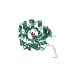
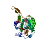
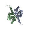
| Structure viewer | Molecule: MolmilJmol/JSmol |
|---|
- Downloads & links
-Download
| PDBx/mmCIF format | 2itg.cif.gz | 45.6 KB | Display | PDBx/mmCIF format |
|---|---|---|---|---|
| PDB format | pdb2itg.ent.gz | 31.7 KB | Display | PDB format |
| PDBx/mmJSON format | 2itg.json.gz | Tree view | PDBx/mmJSON format | |
| Others |  Other downloads Other downloads |
-Validation report
| Summary document | 2itg_validation.pdf.gz | 367.5 KB | Display | wwPDB validaton report |
|---|---|---|---|---|
| Full document | 2itg_full_validation.pdf.gz | 392.2 KB | Display | |
| Data in XML | 2itg_validation.xml.gz | 8.6 KB | Display | |
| Data in CIF | 2itg_validation.cif.gz | 12 KB | Display | |
| Arichive directory | https://data.pdbj.org/pub/pdb/validation_reports/it/2itgftp://data.pdbj.org/pub/pdb/validation_reports/it/2itg | HTTPS FTP |
-Related structure data
| Related structure data |  1itgS S: Starting model for refinement |
|---|---|
| Similar structure data |


-Links
 PDBj
PDBj
- Assembly
| Deposited unit | 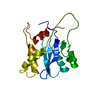
| ||||||||
|---|---|---|---|---|---|---|---|---|---|
| 1 | 
| ||||||||
| Unit cell |
|
-Components
| #1: Protein | Mass: 17921.447 Da / Num. of mol.: 1 / Fragment: CATALYTIC CORE DOMAIN 50 - 212 / Mutation: F185H Source method: isolated from a genetically manipulated source Source: (gene. exp.) Human immunodeficiency virus 1 / Genus: LentivirusDescription: EXPRESSION CLONE FOR CORE, REFER TO PNAS USA, VOL. 90, PP3428-3432, APRIL 1993, AND PNAS USA, VOL. 92, PP.6057-6061, JUNE 1995 Cell line: BL21 / Plasmid: PET-15B / Species (production host): Escherichia coli / Production host:  |
|---|---|
| #2: Water | ChemComp-HOH /  Mass: 18.015 Da / Num. of mol.: 58 / Source method: isolated from a natural source / Formula: H2O Mass: 18.015 Da / Num. of mol.: 58 / Source method: isolated from a natural source / Formula: H2O |
-Experimental details
-Experiment
| Experiment | Method: X-RAY DIFFRACTION / Number of used crystals: 1 |
|---|
- Sample preparation
| Crystal | Density Matthews: 2.58 Å3/Da / Density % sol: 47 % | ||||||||||||||||||||||||||||||||||||||||
|---|---|---|---|---|---|---|---|---|---|---|---|---|---|---|---|---|---|---|---|---|---|---|---|---|---|---|---|---|---|---|---|---|---|---|---|---|---|---|---|---|---|
| Crystal grow | pH: 6.5 Details: 3-9% PEG 8000, 0.4M AMMONIUM SULFATE, 0.1M SODIUM CACODYLATE PH 6.5 | ||||||||||||||||||||||||||||||||||||||||
| Crystal grow | *PLUS Temperature: 19 ℃ / Method: vapor diffusion, sitting drop | ||||||||||||||||||||||||||||||||||||||||
| Components of the solutions | *PLUS
|
-Data collection
| Diffraction | Mean temperature: 298 K |
|---|---|
| Diffraction source | Source: ROTATING ANODE / Type: RIGAKU RUH2R / Wavelength: 1.5418 |
| Detector | Type: RIGAKU / Detector: IMAGE PLATE / Date: Apr 5, 1996 / Details: DOUBLE FOCUSSING MIRRORS |
| Radiation | Monochromatic (M) / Laue (L): M / Scattering type: x-ray |
| Radiation wavelength | Wavelength: 1.5418 Å / Relative weight: 1 |
| Reflection | Resolution: 2.6→10 Å / Num. obs: 5012 / % possible obs: 79.8 % / Observed criterion σ(I): 0 / Redundancy: 2.93 % / Rmerge(I) obs: 0.085 / Net I/σ(I): 8.63 |
| Reflection shell | Resolution: 2.6→2.64 Å / Redundancy: 1.38 % / Rmerge(I) obs: 0.281 / Mean I/σ(I) obs: 1.88 / % possible all: 54.4 |
| Reflection | *PLUS Num. measured all: 14682 |
| Reflection shell | *PLUS % possible obs: 54.4 % |
- Processing
| Software |
| ||||||||||||||||||||||||||||||||||||||||||||||||||||||||||||||||||||||||||||||||||||
|---|---|---|---|---|---|---|---|---|---|---|---|---|---|---|---|---|---|---|---|---|---|---|---|---|---|---|---|---|---|---|---|---|---|---|---|---|---|---|---|---|---|---|---|---|---|---|---|---|---|---|---|---|---|---|---|---|---|---|---|---|---|---|---|---|---|---|---|---|---|---|---|---|---|---|---|---|---|---|---|---|---|---|---|---|---|
| Refinement | Method to determine structure: molecular replacement Starting model: PDB ENTRY 1ITG Resolution: 2.6→10 Å / σ(F): 2
| ||||||||||||||||||||||||||||||||||||||||||||||||||||||||||||||||||||||||||||||||||||
| Displacement parameters | Biso mean: 34.16 Å2 | ||||||||||||||||||||||||||||||||||||||||||||||||||||||||||||||||||||||||||||||||||||
| Refinement step | Cycle: LAST / Resolution: 2.6→10 Å
| ||||||||||||||||||||||||||||||||||||||||||||||||||||||||||||||||||||||||||||||||||||
| Refine LS restraints |
| ||||||||||||||||||||||||||||||||||||||||||||||||||||||||||||||||||||||||||||||||||||
| Software | *PLUS Name: PROLSQ / Classification: refinement | ||||||||||||||||||||||||||||||||||||||||||||||||||||||||||||||||||||||||||||||||||||
| Refinement | *PLUS Rfactor obs: 0.2 | ||||||||||||||||||||||||||||||||||||||||||||||||||||||||||||||||||||||||||||||||||||
| Solvent computation | *PLUS | ||||||||||||||||||||||||||||||||||||||||||||||||||||||||||||||||||||||||||||||||||||
| Displacement parameters | *PLUS |
