- PDB-2ion: Crystal structure of the C-terminal MA3 domain of Pdcd4 (mouse); form2 -
+
Open data
ID or keywords:
Loading...
-
Basic information
Entry
Database: PDB / ID: 2ion
Title
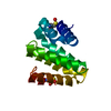
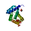
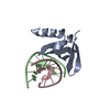
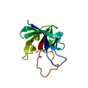
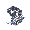
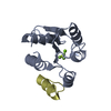
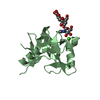
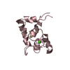
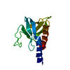
Crystal structure of the C-terminal MA3 domain of Pdcd4 (mouse); form2
Components
Programmed Cell Death 4, Pdcd4
Keywords
ANTITUMOR PROTEIN / alpha-helical
Function / homology
Function and homology information
epithelial to mesenchymal transition involved in cardiac fibroblast development / negative regulation of myofibroblast differentiation / negative regulation of vascular associated smooth muscle cell differentiation / negative regulation of JUN kinase activity / positive regulation of smooth muscle cell apoptotic process / regulation of protein metabolic process / positive regulation of vascular associated smooth muscle cell apoptotic process / negative regulation of vascular associated smooth muscle cell proliferation / positive regulation of endothelial cell apoptotic process / BMP signaling pathway ...epithelial to mesenchymal transition involved in cardiac fibroblast development / negative regulation of myofibroblast differentiation / negative regulation of vascular associated smooth muscle cell differentiation / negative regulation of JUN kinase activity / positive regulation of smooth muscle cell apoptotic process / regulation of protein metabolic process / positive regulation of vascular associated smooth muscle cell apoptotic process / negative regulation of vascular associated smooth muscle cell proliferation / positive regulation of endothelial cell apoptotic process / BMP signaling pathway / negative regulation of cytokine production involved in inflammatory response / positive regulation of non-canonical NF-kappaB signal transduction / positive regulation of inflammatory response / cellular response to lipopolysaccharide / negative regulation of DNA-templated transcription / apoptotic process / negative regulation of apoptotic process / RNA binding / nucleus / cytoplasm / cytosol Similarity search - Function
Programmed cell death protein 4 / Serine Threonine Protein Phosphatase 5, Tetratricopeptide repeat - #180 / Initiation factor eIF-4 gamma, MA3 / MA3 domain / MI domain profile. / Domain in DAP-5, eIF4G, MA-3 and other proteins. / Serine Threonine Protein Phosphatase 5, Tetratricopeptide repeat / Alpha Horseshoe / Armadillo-type fold / Mainly Alpha Similarity search - Domain/homology
In the structure databanks used in Yorodumi, some data are registered as the other names, "COVID-19 virus" and "2019-nCoV". Here are the details of the virus and the list of structure data.
Jan 31, 2019. EMDB accession codes are about to change! (news from PDBe EMDB page)
EMDB accession codes are about to change! (news from PDBe EMDB page)
The allocation of 4 digits for EMDB accession codes will soon come to an end. Whilst these codes will remain in use, new EMDB accession codes will include an additional digit and will expand incrementally as the available range of codes is exhausted. The current 4-digit format prefixed with “EMD-” (i.e. EMD-XXXX) will advance to a 5-digit format (i.e. EMD-XXXXX), and so on. It is currently estimated that the 4-digit codes will be depleted around Spring 2019, at which point the 5-digit format will come into force.
The EM Navigator/Yorodumi systems omit the EMD- prefix.
Related info.:Q: What is EMD? / ID/Accession-code notation in Yorodumi/EM Navigator
Yorodumi is a browser for structure data from EMDB, PDB, SASBDB, etc.
This page is also the successor to EM Navigator detail page, and also detail information page/front-end page for Omokage search.
The word "yorodu" (or yorozu) is an old Japanese word meaning "ten thousand". "mi" (miru) is to see.
Related info.:EMDB / PDB / SASBDB / Comparison of 3 databanks / Yorodumi Search / Aug 31, 2016. New EM Navigator & Yorodumi / Yorodumi Papers / Jmol/JSmol / Function and homology information / Changes in new EM Navigator and Yorodumi
 Movie
Movie Controller

 Yorodumi
 Open data
 Basic information
 Components
 Keywords
 Function and homology information
 X-RAY DIFFRACTION /
  Authors
 Citation
 Structure visualization
 Downloads & links
 Other downloads


 PDBj
PDBj
 Assembly
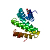


 Mass: 92.094 Da / Num. of mol.: 1 / Source method: obtained synthetically / Formula: C3H8O3
 Mass: 18.015 Da / Num. of mol.: 185 / Source method: isolated from a natural source / Formula: H2O
 Sample preparation
 Processing