 Movie
Movie Controller


+ Open data

- Basic information
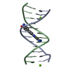
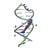
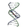
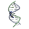
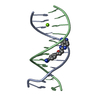
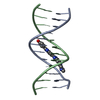
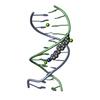
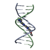
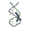
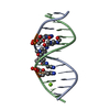
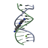
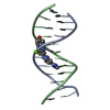
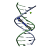
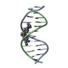
| Entry | Database: PDB / ID: 2i5a | ||||||||||||||||||
|---|---|---|---|---|---|---|---|---|---|---|---|---|---|---|---|---|---|---|---|
| Title | Crystal structure of a DB1055-D(CGCGAATTCGCG)2 complex | ||||||||||||||||||
 Components Components | 5'-D(* Keywords KeywordsDNA / B-type DNA dodecamer with compound DB 1055 | Function / homology | Chem-M1B / DNA / DNA (> 10) |  Function and homology information Function and homology informationMethod |  X-RAY DIFFRACTION / MOLECULAR REPLACEMENT / Resolution: 1.65 Å X-RAY DIFFRACTION / MOLECULAR REPLACEMENT / Resolution: 1.65 Å  Authors AuthorsNeidle, S. / Lee, M.P.H. |  CitationJournal: To be Published CitationJournal: To be PublishedTitle: Crystal structure of a DB1055-D(CGCGAATTCGCG)2 complex Authors: Neidle, S. / Lee, M.P.H. History |
|
- Structure visualization
| Structure viewer | Molecule: MolmilJmol/JSmol |
|---|
- Downloads & links
-Download
| PDBx/mmCIF format | 2i5a.cif.gz | 26.4 KB | Display | PDBx/mmCIF format |
|---|---|---|---|---|
| PDB format | pdb2i5a.ent.gz | 16.2 KB | Display | PDB format |
| PDBx/mmJSON format | 2i5a.json.gz | Tree view | PDBx/mmJSON format | |
| Others |  Other downloads Other downloads |
-Validation report
| Arichive directory | https://data.pdbj.org/pub/pdb/validation_reports/i5/2i5aftp://data.pdbj.org/pub/pdb/validation_reports/i5/2i5a | HTTPS FTP |
|---|
-Related structure data
| Related structure data |  2dbeS S: Starting model for refinement |
|---|---|
| Similar structure data |


-Links
 PDBj
PDBj


- Assembly
| Deposited unit | 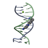
| ||||||||
|---|---|---|---|---|---|---|---|---|---|
| 1 |
| ||||||||
| Unit cell |
|
-Components
| #1: DNA chain | Mass: 3663.392 Da / Num. of mol.: 2 / Source method: obtained synthetically #2: Chemical | ChemComp-MG / |   Mass: 24.305 Da / Num. of mol.: 1 / Source method: obtained synthetically / Formula: Mg Mass: 24.305 Da / Num. of mol.: 1 / Source method: obtained synthetically / Formula: Mg#3: Chemical | ChemComp-M1B / | 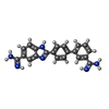  Mass: 354.408 Da / Num. of mol.: 1 / Source method: obtained synthetically / Formula: C21H18N6 Mass: 354.408 Da / Num. of mol.: 1 / Source method: obtained synthetically / Formula: C21H18N6#4: Water | ChemComp-HOH / |  Mass: 18.015 Da / Num. of mol.: 84 / Source method: isolated from a natural source / Formula: H2O Mass: 18.015 Da / Num. of mol.: 84 / Source method: isolated from a natural source / Formula: H2O |
|---|
-Experimental details
-Experiment
| Experiment | Method: X-RAY DIFFRACTION / Number of used crystals: 1 |
|---|
- Sample preparation
| Crystal | Density Matthews: 2.07 Å3/Da / Density % sol: 40.56 % | ||||||||||||||||||||||||||||||||||||
|---|---|---|---|---|---|---|---|---|---|---|---|---|---|---|---|---|---|---|---|---|---|---|---|---|---|---|---|---|---|---|---|---|---|---|---|---|---|
| Crystal grow | pH: 7 Details: MAGNESIUM CHLORIDE, DNA DODECAMER, MPD, COMPOUND DB 1055, SODIUM CACODYLATE BUFFER, pH 7, VAPOR DIFFUSION, HANGING DROP, pH 7.00 | ||||||||||||||||||||||||||||||||||||
| Components of the solutions |
|
-Data collection
| Diffraction | Mean temperature: 105 K |
|---|---|
| Diffraction source | Source: ROTATING ANODE / Type: RIGAKU RU200 / Wavelength: 1.5418 |
| Detector | Type: RIGAKU RAXIS IV / Detector: IMAGE PLATE / Date: Jul 17, 2006 / Details: OSMIC FOCUSING MIRROR SYSTEM |
| Radiation | Monochromator: NI FILTER / Protocol: SINGLE WAVELENGTH / Monochromatic (M) / Laue (L): M / Scattering type: x-ray |
| Radiation wavelength | Wavelength: 1.5418 Å / Relative weight: 1 |
| Reflection | Resolution: 1.65→25 Å / Num. obs: 7580 / % possible obs: 93.1 % / Observed criterion σ(I): 0 / Redundancy: 2.91 % / Rmerge(I) obs: 0.035 / Net I/σ(I): 33.42 |
| Reflection shell | Resolution: 1.65→1.71 Å / Rmerge(I) obs: 0.219 / Mean I/σ(I) obs: 4.8 / % possible all: 90.7 |
- Processing
| Software |
| |||||||||||||||||||||||||||||||||
|---|---|---|---|---|---|---|---|---|---|---|---|---|---|---|---|---|---|---|---|---|---|---|---|---|---|---|---|---|---|---|---|---|---|---|
| Refinement | Method to determine structure: MOLECULAR REPLACEMENT Starting model: PDB ENTRY 2DBE Resolution: 1.65→8 Å / Num. parameters: 2359 / Num. restraintsaints: 2359 / Cross valid method: THROUGHOUT / σ(F): 2 / Stereochemistry target values: ENGH & HUBER
| |||||||||||||||||||||||||||||||||
| Solvent computation | Solvent model: MOEWS & KRETSINGER, J.MOL.BIOL.91(1973)201-22 | |||||||||||||||||||||||||||||||||
| Refine analyze | Num. disordered residues: 0 / Occupancy sum hydrogen: 0 / Occupancy sum non hydrogen: 598 | |||||||||||||||||||||||||||||||||
| Refinement step | Cycle: LAST / Resolution: 1.65→8 Å
| |||||||||||||||||||||||||||||||||
| Refine LS restraints |
|
