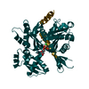
- PDB-2i51: CRYSTAL STRUCTURE OF a pyridoxamine 5'-phosphate oxidase-related,... -
+
Open data
ID or keywords:
Loading...
-
Basic information
Entry
Database: PDB / ID: 2i51
Title
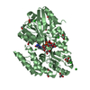
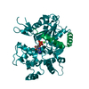
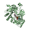
CRYSTAL STRUCTURE OF a pyridoxamine 5'-phosphate oxidase-related, FMN binding protein (NPUN_F5749) FROM NOSTOC PUNCTIFORME PCC 73102 AT 1.40 A RESOLUTION
Components
Uncharacterized conserved protein of COG5135
Keywords
FLAVOPROTEIN / PYRIDOXAMINE 5'-PHOSPHATE OXIDASE-RELATED PROTEIN / STRUCTURAL GENOMICS / JOINT CENTER FOR STRUCTURAL GENOMICS / JCSG / PROTEIN STRUCTURE INITIATIVE / PSI-2
BIOMOLECULE: 1 THIS ENTRY CONTAINS THE CRYSTALLOGRAPHIC ASYMMETRIC UNIT WHICH CONSISTS OF 2 CHAIN(S) ...BIOMOLECULE: 1 THIS ENTRY CONTAINS THE CRYSTALLOGRAPHIC ASYMMETRIC UNIT WHICH CONSISTS OF 2 CHAIN(S). SEE REMARK 350 FOR INFORMATION ON GENERATING THE BIOLOGICAL MOLECULE(S). SIZE EXCLUSION CHROMATOGRAPHY DATA SUPPORTS THE ASSIGNMENT OF A DIMER AS A BIOLOGICALLY SIGNIFICANT OLIGOMERIZATION STATE.
Remark 999
SEQUENCE THE CONSTRUCT WAS EXPRESSED WITH A PURIFICATION TAG MGSDKIHHHHHHENLYFQG. THE TAG WAS ...SEQUENCE THE CONSTRUCT WAS EXPRESSED WITH A PURIFICATION TAG MGSDKIHHHHHHENLYFQG. THE TAG WAS REMOVED WITH TEV PROTEASE LEAVING ONLY A GLYCINE (0) FOLLOWED BY THE TARGET SEQUENCE. A suitable database reference was not available at the time of processing.
Monochromator: Single crystal Si(111) bent monochromator (horizontal focusing) Protocol: MAD / Monochromatic (M) / Laue (L): M / Scattering type: x-ray
Radiation wavelength
ID
Wavelength (Å)
Relative weight
1
0.978489
1
2
0.979264
1
3
0.911617
1
Reflection
Resolution: 1.397→29.386 Å / Num. obs: 96246 / % possible obs: 99.7 % / Redundancy: 5.2 % / Biso Wilson estimate: 15.91 Å2 / Rmerge(I) obs: 0.113 / Rsym value: 0.113 / Net I/σ(I): 3.5
Reflection shell
Rmerge(I) obs: 0.01 / Diffraction-ID: 2
Resolution (Å)
Redundancy (%)
Mean I/σ(I) obs
Num. measured all
Num. unique all
Rsym value
% possible all
1.4-1.44
4.6
0.7
31896
6910
0.01016
98
1.44-1.48
4.7
0.8
32178
6899
0.874
100
1.48-1.52
4.7
1
31398
6716
0.758
100
1.52-1.57
4.7
1.2
30561
6513
0.604
100
1.57-1.62
4.7
1.5
29438
6277
0.488
100
1.62-1.67
4.7
1.5
28836
6119
0.395
100
1.67-1.74
4.7
2.3
27948
5939
0.317
99.9
1.74-1.81
4.7
2.9
26601
5653
0.254
100
1.81-1.89
4.7
3.9
25893
5508
0.184
100
1.89-1.98
4.7
4.7
24472
5199
0.142
99.8
1.98-2.09
4.7
6.2
23255
4944
0.107
99.8
2.09-2.21
6.4
3.6
30475
4749
0.157
100
2.21-2.37
6.4
4.4
28711
4480
0.135
100
2.37-2.56
6.4
4.5
26467
4135
0.126
100
2.56-2.8
6.3
5.1
24064
3823
0.116
100
2.8-3.13
6.1
5.4
21322
3503
0.109
99.9
3.13-3.61
5.8
6.2
17562
3052
0.092
98.7
3.61-4.43
6.4
7.8
16838
2636
0.07
99.6
4.43-6.26
6.1
8.2
12415
2049
0.067
99.2
6.26-29.44
5.7
9.3
6520
1142
0.066
94.1
-
Phasing
Phasing
Method: MAD
-
Processing
Software
Name
Version
Classification
NB
MolProbity
3beta29
modelbuilding
SHELX
phasing
REFMAC
5.2.0019
refinement
SCALA
datascaling
PDB_EXTRACT
2
dataextraction
MOSFLM
datareduction
CCP4
(SCALA)
datascaling
autoSHARP
phasing
Refinement
Method to determine structure: MAD / Resolution: 1.4→29.386 Å / Cor.coef. Fo:Fc: 0.973 / Cor.coef. Fo:Fc free: 0.963 / SU B: 1.907 / SU ML: 0.038 / TLS residual ADP flag: LIKELY RESIDUAL / Cross valid method: THROUGHOUT / σ(F): 0 / ESU R: 0.052 / ESU R Free: 0.054 Stereochemistry target values: MAXIMUM LIKELIHOOD WITH PHASES Details: 1. HYDROGENS HAVE BEEN ADDED IN THE RIDING POSITIONS 2. A MET-INHIBITION PROTOCOL WAS USED FOR SELENOMETHIONINE INCORPORATION DURING PROTEIN EXPRESSION. THE OCCUPANCY OF THE SE ATOMS IN THE ...Details: 1. HYDROGENS HAVE BEEN ADDED IN THE RIDING POSITIONS 2. A MET-INHIBITION PROTOCOL WAS USED FOR SELENOMETHIONINE INCORPORATION DURING PROTEIN EXPRESSION. THE OCCUPANCY OF THE SE ATOMS IN THE MSE RESIDUES WAS REDUCED TO 0.75 TO ACCOUNT FOR THE REDUCED SCATTERING POWER DUE TO PARTIAL S-MET INCORPORATION. 3. ATOM RECORD CONTAINS RESIDUAL B FACTORS ONLY. 4. RESIDUES B134-137 ARE DISORDERED AND WERE NOT MODELED. 5. FMN MODELED BASED ON PROPOSED FUNCTION AND DENSITY. 6. ETHYLENE GLYCOL AND GLYCEROL MODELED BASED ON CRYSTALLIAZTION CONDITIONS. 7. THERE IS UNMODELED DENSITY NEAR A45, A115, AND B153.
Rfactor
Num. reflection
% reflection
Selection details
Rfree
0.188
4853
5 %
RANDOM
Rwork
0.165
-
-
-
obs
0.166
96170
99.59 %
-
Solvent computation
Ion probe radii: 0.8 Å / Shrinkage radii: 0.8 Å / VDW probe radii: 1.2 Å / Solvent model: MASK
Displacement parameters
Biso mean: 16.71 Å2
Baniso -1
Baniso -2
Baniso -3
1-
-0.01 Å2
0 Å2
0 Å2
2-
-
0.45 Å2
0 Å2
3-
-
-
-0.44 Å2
Refinement step
Cycle: LAST / Resolution: 1.4→29.386 Å
Protein
Nucleic acid
Ligand
Solvent
Total
Num. atoms
3120
0
148
359
3627
Refine LS restraints
Refine-ID
Type
Dev ideal
Dev ideal target
Number
X-RAY DIFFRACTION
r_bond_refined_d
0.016
0.022
3453
X-RAY DIFFRACTION
r_bond_other_d
0.002
0.02
2455
X-RAY DIFFRACTION
r_angle_refined_deg
1.637
1.979
4706
X-RAY DIFFRACTION
r_angle_other_deg
0.974
3
5908
X-RAY DIFFRACTION
r_dihedral_angle_1_deg
5.541
5
414
X-RAY DIFFRACTION
r_dihedral_angle_2_deg
36.686
22.989
174
X-RAY DIFFRACTION
r_dihedral_angle_3_deg
12.16
15
519
X-RAY DIFFRACTION
r_dihedral_angle_4_deg
15.905
15
36
X-RAY DIFFRACTION
r_chiral_restr
0.107
0.2
480
X-RAY DIFFRACTION
r_gen_planes_refined
0.012
0.02
3799
X-RAY DIFFRACTION
r_gen_planes_other
0.001
0.02
717
X-RAY DIFFRACTION
r_nbd_refined
0.213
0.2
631
X-RAY DIFFRACTION
r_nbd_other
0.227
0.2
2783
X-RAY DIFFRACTION
r_nbtor_refined
0.189
0.2
1636
X-RAY DIFFRACTION
r_nbtor_other
0.086
0.2
1895
X-RAY DIFFRACTION
r_xyhbond_nbd_refined
0.166
0.2
288
X-RAY DIFFRACTION
r_symmetry_vdw_refined
0.31
0.2
17
X-RAY DIFFRACTION
r_symmetry_vdw_other
0.331
0.2
46
X-RAY DIFFRACTION
r_symmetry_hbond_refined
0.193
0.2
9
X-RAY DIFFRACTION
r_mcbond_it
2.135
3
2069
X-RAY DIFFRACTION
r_mcbond_other
0.506
3
768
X-RAY DIFFRACTION
r_mcangle_it
3.065
5
3251
X-RAY DIFFRACTION
r_scbond_it
4.321
8
1684
X-RAY DIFFRACTION
r_scangle_it
6.091
11
1440
LS refinement shell
Resolution: 1.4→1.436 Å / Total num. of bins used: 20
Rfactor
Num. reflection
% reflection
Rfree
0.347
318
-
Rwork
0.323
6569
-
obs
-
6887
97.94 %
Refinement TLS params.
Method: refined / Refine-ID: X-RAY DIFFRACTION
ID
L11 (°2)
L12 (°2)
L13 (°2)
L22 (°2)
L23 (°2)
L33 (°2)
S11 (Å °)
S12 (Å °)
S13 (Å °)
S21 (Å °)
S22 (Å °)
S23 (Å °)
S31 (Å °)
S32 (Å °)
S33 (Å °)
T11 (Å2)
T12 (Å2)
T13 (Å2)
T22 (Å2)
T23 (Å2)
T33 (Å2)
Origin x (Å)
Origin y (Å)
Origin z (Å)
1
0.2245
0.2098
0.0872
0.5216
0.3082
1.0564
-0.0455
0.0565
0.0301
-0.0911
-0.0129
0.0541
-0.0817
0.0031
0.0585
-0.0376
0.0012
-0.0072
-0.046
0.0014
-0.0389
48.0279
62.1066
4.2312
2
0.4849
0.0901
0.1257
0.6878
0.4465
1.139
-0.0204
-0.0764
0.062
0.0024
-0.0546
0.1077
-0.1309
-0.039
0.075
-0.0276
-0.0009
0.0093
-0.0591
-0.0142
-0.0234
47.2543
73.0979
24.3294
Refinement TLS group
Refine-ID: X-RAY DIFFRACTION / Selection: ALL
ID
Refine TLS-ID
Auth asym-ID
Label asym-ID
Auth seq-ID
Label seq-ID
1
1
A
A
1 - 194
2 - 195
2
2
B
B
0 - 194
1 - 195
+
About Yorodumi
-
News
-
Feb 9, 2022. New format data for meta-information of EMDB entries
New format data for meta-information of EMDB entries
Version 3 of the EMDB header file is now the official format.
The previous official version 1.9 will be removed from the archive.
In the structure databanks used in Yorodumi, some data are registered as the other names, "COVID-19 virus" and "2019-nCoV". Here are the details of the virus and the list of structure data.
Jan 31, 2019. EMDB accession codes are about to change! (news from PDBe EMDB page)
EMDB accession codes are about to change! (news from PDBe EMDB page)
The allocation of 4 digits for EMDB accession codes will soon come to an end. Whilst these codes will remain in use, new EMDB accession codes will include an additional digit and will expand incrementally as the available range of codes is exhausted. The current 4-digit format prefixed with “EMD-” (i.e. EMD-XXXX) will advance to a 5-digit format (i.e. EMD-XXXXX), and so on. It is currently estimated that the 4-digit codes will be depleted around Spring 2019, at which point the 5-digit format will come into force.
The EM Navigator/Yorodumi systems omit the EMD- prefix.
Related info.:Q: What is EMD? / ID/Accession-code notation in Yorodumi/EM Navigator
Yorodumi is a browser for structure data from EMDB, PDB, SASBDB, etc.
This page is also the successor to EM Navigator detail page, and also detail information page/front-end page for Omokage search.
The word "yorodu" (or yorozu) is an old Japanese word meaning "ten thousand". "mi" (miru) is to see.
Related info.:EMDB / PDB / SASBDB / Comparison of 3 databanks / Yorodumi Search / Aug 31, 2016. New EM Navigator & Yorodumi / Yorodumi Papers / Jmol/JSmol / Function and homology information / Changes in new EM Navigator and Yorodumi
 Movie
Movie Controller

 Yorodumi
 Open data
 Basic information
 Components
 Keywords
 Function and homology information
 Nostoc punctiforme (bacteria)
 X-RAY DIFFRACTION /
  Authors
 Citation
 Structure visualization
 Downloads & links
 Other downloads
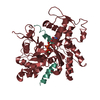


 PDBj
PDBj Assembly

 Mass: 456.344 Da / Num. of mol.: 2 / Source method: obtained synthetically / Formula: C17H21N4O9P
 Mass: 62.068 Da / Num. of mol.: 20 / Source method: obtained synthetically / Formula: C2H6O2
 Mass: 92.094 Da / Num. of mol.: 1 / Source method: obtained synthetically / Formula: C3H8O3
 Mass: 18.015 Da / Num. of mol.: 359 / Source method: isolated from a natural source / Formula: H2O
 Sample preparation
 Processing