 Movie
Movie Controller

[English] 日本語
 Yorodumi
- PDB-2hzf: Crystal structures of a poxviral glutaredoxin in the oxidized and... -
+ Open data

- Basic information
| Entry | Database: PDB / ID: 2hzf | ||||||
|---|---|---|---|---|---|---|---|
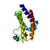
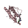
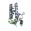
| Title | Crystal structures of a poxviral glutaredoxin in the oxidized and reduced states show redox-correlated structural changes | ||||||
 Components Components | Glutaredoxin-1 | ||||||
 Keywords Keywords | ELECTRON TRANSPORT / OXIDOREDUCTASE / thioredoxin fold | ||||||
| Function / homology |  Function and homology information Function and homology information | ||||||
| Biological species |  Ectromelia virus Ectromelia virus | ||||||
| Method |  X-RAY DIFFRACTION / SYNCHROTRON / MOLECULAR REPLACEMENT / Resolution: 1.8 Å X-RAY DIFFRACTION / SYNCHROTRON / MOLECULAR REPLACEMENT / Resolution: 1.8 Å | ||||||
 Authors Authors | Bacik, J.P. / Hazes, B. | ||||||
 Citation Citation | Journal: J.Mol.Biol. / Year: 2007 Title: Crystal Structures of a Poxviral Glutaredoxin in the Oxidized and Reduced States Show Redox-correlated Structural Changes. Authors: Bacik, J.P. / Hazes, B. | ||||||
| History |
|
- Structure visualization
| Structure viewer | Molecule: MolmilJmol/JSmol |
|---|
- Downloads & links
-Download
| PDBx/mmCIF format | 2hzf.cif.gz | 58.6 KB | Display | PDBx/mmCIF format |
|---|---|---|---|---|
| PDB format | pdb2hzf.ent.gz | 42.9 KB | Display | PDB format |
| PDBx/mmJSON format | 2hzf.json.gz | Tree view | PDBx/mmJSON format | |
| Others |  Other downloads Other downloads |
-Validation report
| Arichive directory | https://data.pdbj.org/pub/pdb/validation_reports/hz/2hzfftp://data.pdbj.org/pub/pdb/validation_reports/hz/2hzf | HTTPS FTP |
|---|
-Related structure data


-Links
 PDBj
PDBj- Assembly
| Deposited unit | 
| ||||||||||||
|---|---|---|---|---|---|---|---|---|---|---|---|---|---|
| 1 | 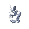
| ||||||||||||
| 2 | 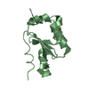
| ||||||||||||
| Unit cell |
| ||||||||||||
| Components on special symmetry positions |
|
-Components
| #1: Protein | Mass: 13160.028 Da / Num. of mol.: 2 Source method: isolated from a genetically manipulated source Source: (gene. exp.) Ectromelia virus / Genus: Orthopoxvirus / Strain: Moscow / Gene: EVM053 / Production host:  References: UniProt: Q8JLF5, glutathione dehydrogenase (ascorbate) #2: Water | ChemComp-HOH / |  Mass: 18.015 Da / Num. of mol.: 148 / Source method: isolated from a natural source / Formula: H2O Mass: 18.015 Da / Num. of mol.: 148 / Source method: isolated from a natural source / Formula: H2OHas protein modification | Y | |
|---|
-Experimental details
-Experiment
| Experiment | Method: X-RAY DIFFRACTION / Number of used crystals: 1 |
|---|
- Sample preparation
| Crystal | Density Matthews: 2.16 Å3/Da / Density % sol: 43.02 % |
|---|---|
| Crystal grow | Temperature: 295 K / Method: vapor diffusion, hanging drop / pH: 6 Details: 20 % MPD, 0.1 M NaCacodylate pH 6.0, 5 mM DTT, 10 mM Tris pH 8.0, 100 mM sodium chloride, VAPOR DIFFUSION, HANGING DROP, temperature 295K |
-Data collection
| Diffraction | Mean temperature: 100 K |
|---|---|
| Diffraction source | Source: SYNCHROTRON / Site: ALS  / Beamline: 8.3.1 / Wavelength: 1.1 Å / Beamline: 8.3.1 / Wavelength: 1.1 Å |
| Detector | Type: ADSC QUANTUM 210 / Detector: CCD / Date: Jan 14, 2005 |
| Radiation | Monochromator: double crystal / Protocol: SINGLE WAVELENGTH / Monochromatic (M) / Laue (L): M / Scattering type: x-ray |
| Radiation wavelength | Wavelength: 1.1 Å / Relative weight: 1 |
| Reflection | Resolution: 1.8→36.2 Å / Num. all: 20896 / Num. obs: 20896 / % possible obs: 96.7 % / Observed criterion σ(F): 3.24 / Observed criterion σ(I): 1.34 |
| Reflection shell | Resolution: 1.8→1.9 Å / % possible all: 88 |
- Processing
| Software |
| ||||||||||||||||||||||||||||||||||||||||||||||||||||||||||||||||||||||||||||||||||||||||||||||||||||||||||||||||||||||||||||||||||||||||||||||||||||||||||||||||||||||||||
|---|---|---|---|---|---|---|---|---|---|---|---|---|---|---|---|---|---|---|---|---|---|---|---|---|---|---|---|---|---|---|---|---|---|---|---|---|---|---|---|---|---|---|---|---|---|---|---|---|---|---|---|---|---|---|---|---|---|---|---|---|---|---|---|---|---|---|---|---|---|---|---|---|---|---|---|---|---|---|---|---|---|---|---|---|---|---|---|---|---|---|---|---|---|---|---|---|---|---|---|---|---|---|---|---|---|---|---|---|---|---|---|---|---|---|---|---|---|---|---|---|---|---|---|---|---|---|---|---|---|---|---|---|---|---|---|---|---|---|---|---|---|---|---|---|---|---|---|---|---|---|---|---|---|---|---|---|---|---|---|---|---|---|---|---|---|---|---|---|---|---|---|
| Refinement | Method to determine structure: MOLECULAR REPLACEMENT / Resolution: 1.8→19.76 Å / Cor.coef. Fo:Fc: 0.957 / Cor.coef. Fo:Fc free: 0.936 / Cross valid method: THROUGHOUT / σ(F): 0 / ESU R: 0.132 / ESU R Free: 0.126 / Stereochemistry target values: MAXIMUM LIKELIHOOD / Details: HYDROGENS HAVE BEEN ADDED IN THE RIDING POSITIONS
| ||||||||||||||||||||||||||||||||||||||||||||||||||||||||||||||||||||||||||||||||||||||||||||||||||||||||||||||||||||||||||||||||||||||||||||||||||||||||||||||||||||||||||
| Solvent computation | Ion probe radii: 0.8 Å / Shrinkage radii: 0.8 Å / VDW probe radii: 1.4 Å / Solvent model: BABINET MODEL WITH MASK | ||||||||||||||||||||||||||||||||||||||||||||||||||||||||||||||||||||||||||||||||||||||||||||||||||||||||||||||||||||||||||||||||||||||||||||||||||||||||||||||||||||||||||
| Displacement parameters | Biso mean: 18.256 Å2
| ||||||||||||||||||||||||||||||||||||||||||||||||||||||||||||||||||||||||||||||||||||||||||||||||||||||||||||||||||||||||||||||||||||||||||||||||||||||||||||||||||||||||||
| Refinement step | Cycle: LAST / Resolution: 1.8→19.76 Å
| ||||||||||||||||||||||||||||||||||||||||||||||||||||||||||||||||||||||||||||||||||||||||||||||||||||||||||||||||||||||||||||||||||||||||||||||||||||||||||||||||||||||||||
| Refine LS restraints |
| ||||||||||||||||||||||||||||||||||||||||||||||||||||||||||||||||||||||||||||||||||||||||||||||||||||||||||||||||||||||||||||||||||||||||||||||||||||||||||||||||||||||||||
| LS refinement shell | Highest resolution: 1.8 Å / Total num. of bins used: 20 /
|
