 Movie
Movie Controller

[English] 日本語
 Yorodumi
- PDB-2huf: Crystal structure of Aedes aegypti alanine glyoxylate aminotransferase -
+ Open data

- Basic information
| Entry | Database: PDB / ID: 2huf | ||||||
|---|---|---|---|---|---|---|---|
| Title | Crystal structure of Aedes aegypti alanine glyoxylate aminotransferase | ||||||
 Components Components | Alanine glyoxylate aminotransferase | ||||||
 Keywords Keywords | TRANSFERASE / Alpha and beta protein / PLP-dependent transferase | ||||||
| Function / homology |  Function and homology information Function and homology informationalanine-glyoxylate transaminase / L-serine:pyruvate transaminase activity / : / L-alanine:glyoxylate transaminase activity / glyoxylate catabolic process / pyridoxal phosphate binding / peroxisome Similarity search - Function | ||||||
| Biological species |  | ||||||
| Method |  X-RAY DIFFRACTION / SYNCHROTRON / MOLECULAR REPLACEMENT / Resolution: 1.75 Å X-RAY DIFFRACTION / SYNCHROTRON / MOLECULAR REPLACEMENT / Resolution: 1.75 Å | ||||||
 Authors Authors | Han, Q. / Robinson, H. / Gao, Y.G. / Vogelaar, N. / Wilson, S.R. / Rizzi, M. / Li, J. | ||||||
 Citation Citation | Journal: J.Biol.Chem. / Year: 2006 Title: Crystal Structures of Aedes aegypti Alanine Glyoxylate Aminotransferase. Authors: Han, Q. / Robinson, H. / Gao, Y.G. / Vogelaar, N. / Wilson, S.R. / Rizzi, M. / Li, J. | ||||||
| History |
|
- Structure visualization
| Structure viewer | Molecule: MolmilJmol/JSmol |
|---|
- Downloads & links
-Download
| PDBx/mmCIF format | 2huf.cif.gz | 168.8 KB | Display | PDBx/mmCIF format |
|---|---|---|---|---|
| PDB format | pdb2huf.ent.gz | 131.8 KB | Display | PDB format |
| PDBx/mmJSON format | 2huf.json.gz | Tree view | PDBx/mmJSON format | |
| Others |  Other downloads Other downloads |
-Validation report
| Arichive directory | https://data.pdbj.org/pub/pdb/validation_reports/hu/2hufftp://data.pdbj.org/pub/pdb/validation_reports/hu/2huf | HTTPS FTP |
|---|
-Related structure data
| Related structure data |  2huiC 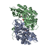 2huuC 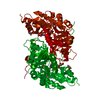 1h0cS C: citing same article ( S: Starting model for refinement |
|---|---|
| Similar structure data |


-Links
 PDBj
PDBj- Assembly
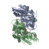
| Deposited unit | 
| ||||||||
|---|---|---|---|---|---|---|---|---|---|
| 1 |
| ||||||||
| Unit cell |
|
-Components
| #1: Protein | Mass: 43390.840 Da / Num. of mol.: 2 Source method: isolated from a genetically manipulated source Source: (gene. exp.)  Spodoptera frugiperda (fall armyworm) / Strain (production host): SF9 Spodoptera frugiperda (fall armyworm) / Strain (production host): SF9References: UniProt: Q3LSM4, alanine-glyoxylate transaminase #2: Chemical | 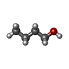  Mass: 74.122 Da / Num. of mol.: 2 / Source method: obtained synthetically / Formula: C4H10O Mass: 74.122 Da / Num. of mol.: 2 / Source method: obtained synthetically / Formula: C4H10O#3: Water | ChemComp-HOH / |  Mass: 18.015 Da / Num. of mol.: 520 / Source method: isolated from a natural source / Formula: H2O Mass: 18.015 Da / Num. of mol.: 520 / Source method: isolated from a natural source / Formula: H2O |
|---|
-Experimental details
-Experiment
| Experiment | Method: X-RAY DIFFRACTION / Number of used crystals: 1 |
|---|
- Sample preparation
| Crystal | Density Matthews: 2.5 Å3/Da / Density % sol: 50.89 % |
|---|---|
| Crystal grow | Temperature: 277 K / Method: vapor diffusion, hanging drop / pH: 6.5 Details: 12% PEG 8000, 0.13 M magnesium acetate, 4% butanol, 0.1 M cacodylic acid, pH 6.5, VAPOR DIFFUSION, HANGING DROP, temperature 277K |
-Data collection
| Diffraction | Mean temperature: 200 K |
|---|---|
| Diffraction source | Source: SYNCHROTRON / Site: NSLS  / Beamline: X25 / Wavelength: 0.9795 Å / Beamline: X25 / Wavelength: 0.9795 Å |
| Detector | Type: ADSC QUANTUM 315 / Detector: CCD / Date: Jun 21, 2006 |
| Radiation | Monochromator: Si(111) or (220) / Protocol: SINGLE WAVELENGTH / Monochromatic (M) / Laue (L): M / Scattering type: x-ray |
| Radiation wavelength | Wavelength: 0.9795 Å / Relative weight: 1 |
| Reflection | Resolution: 1.75→30 Å / Num. obs: 84787 / % possible obs: 100 % / Observed criterion σ(F): 0 / Observed criterion σ(I): 0 / Redundancy: 11 % / Rmerge(I) obs: 0.081 |
| Reflection shell | Resolution: 1.75→1.81 Å / % possible all: 100 |
- Processing
| Software |
| ||||||||||||||||||||||||||||||||||||||||||||||||||||||||||||||||||||||||||||||||||||||||||
|---|---|---|---|---|---|---|---|---|---|---|---|---|---|---|---|---|---|---|---|---|---|---|---|---|---|---|---|---|---|---|---|---|---|---|---|---|---|---|---|---|---|---|---|---|---|---|---|---|---|---|---|---|---|---|---|---|---|---|---|---|---|---|---|---|---|---|---|---|---|---|---|---|---|---|---|---|---|---|---|---|---|---|---|---|---|---|---|---|---|---|---|
| Refinement | Method to determine structure: MOLECULAR REPLACEMENT Starting model: PDB ENTRY 1h0c Resolution: 1.75→29.2 Å / Cor.coef. Fo:Fc: 0.959 / Cor.coef. Fo:Fc free: 0.944 / SU B: 1.903 / SU ML: 0.063 / Isotropic thermal model: Isotropic / Cross valid method: THROUGHOUT / σ(F): 0 / ESU R: 0.106 / ESU R Free: 0.103 / Stereochemistry target values: MAXIMUM LIKELIHOOD / Details: HYDROGENS HAVE BEEN ADDED IN THE RIDING POSITIONS
| ||||||||||||||||||||||||||||||||||||||||||||||||||||||||||||||||||||||||||||||||||||||||||
| Solvent computation | Ion probe radii: 0.8 Å / Shrinkage radii: 0.8 Å / VDW probe radii: 1.2 Å / Solvent model: MASK | ||||||||||||||||||||||||||||||||||||||||||||||||||||||||||||||||||||||||||||||||||||||||||
| Displacement parameters | Biso mean: 18.577 Å2
| ||||||||||||||||||||||||||||||||||||||||||||||||||||||||||||||||||||||||||||||||||||||||||
| Refinement step | Cycle: LAST / Resolution: 1.75→29.2 Å
| ||||||||||||||||||||||||||||||||||||||||||||||||||||||||||||||||||||||||||||||||||||||||||
| Refine LS restraints |
| ||||||||||||||||||||||||||||||||||||||||||||||||||||||||||||||||||||||||||||||||||||||||||
| LS refinement shell | Resolution: 1.751→1.796 Å / Total num. of bins used: 20
|
