 Movie
Movie Controller

[English] 日本語
 Yorodumi
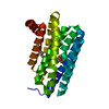
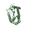
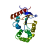
- PDB-2hrx: X-Ray Crystal Structure of Protein DIP2367 from Corynebacterium d... -
+ Open data

- Basic information
| Entry | Database: PDB / ID: 2hrx | ||||||
|---|---|---|---|---|---|---|---|
| Title | X-Ray Crystal Structure of Protein DIP2367 from Corynebacterium diphtheriae. Northeast Structural Genomics Consortium Target CdR13. | ||||||
 Components Components | Hypothetical protein | ||||||
 Keywords Keywords | STRUCTURAL GENOMICS / UNKNOWN FUNCTION / CDR13 NESG Corynebacterium diphtheriae / PSI-2 / Protein Structure Initiative / Northeast Structural Genomics Consortium | ||||||
| Function / homology | VC0467-like / VC0467-like / Protein of unknown function UPF0301 / Uncharacterized ACR, COG1678 / 3-Layer(aba) Sandwich / cytosol / Alpha Beta / YqgE/AlgH family protein Function and homology information Function and homology information | ||||||
| Biological species |  Corynebacterium diphtheriae (bacteria) Corynebacterium diphtheriae (bacteria) | ||||||
| Method |  X-RAY DIFFRACTION / SYNCHROTRON / SAD / Resolution: 1.9 Å X-RAY DIFFRACTION / SYNCHROTRON / SAD / Resolution: 1.9 Å | ||||||
 Authors Authors | Kuzin, A.P. / Su, M. / Jayaraman, S. / Ho, C.K. / Janjua, H. / Cunningham, K. / Ma, L.C. / Xiao, R. / Liu, J. / Baran, M. ...Kuzin, A.P. / Su, M. / Jayaraman, S. / Ho, C.K. / Janjua, H. / Cunningham, K. / Ma, L.C. / Xiao, R. / Liu, J. / Baran, M. / Acton, T.B. / Rost, B. / Montelione, G.T. / Tong, L. / Hunt, J.F. / Northeast Structural Genomics Consortium (NESG) | ||||||
 Citation Citation | Journal: TO BE PUBLISHED Title: Three dimensional structure of conserved hypothetical protein from Corynebacterium diphtheriae at the resolution 1.9 A. Northeast Structural Genomics Consortium target CdR13. Authors: Kuzin, A.P. / Su, M. / Jayaraman, S. / Ho, C.K. / Janjua, H. / Cunningham, K. / Ma, L.C. / Xiao, R. / Liu, J. / Baran, M. / Acton, T.B. / Rost, B. / Montelione, G.T. / Tong, L. / Hunt, J.F. | ||||||
| History |
|
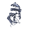
- Structure visualization
| Structure viewer | Molecule: MolmilJmol/JSmol |
|---|
- Downloads & links
-Download
| PDBx/mmCIF format | 2hrx.cif.gz | 49.2 KB | Display | PDBx/mmCIF format |
|---|---|---|---|---|
| PDB format | pdb2hrx.ent.gz | 38.3 KB | Display | PDB format |
| PDBx/mmJSON format | 2hrx.json.gz | Tree view | PDBx/mmJSON format | |
| Others |  Other downloads Other downloads |
-Validation report
| Summary document | 2hrx_validation.pdf.gz | 427 KB | Display | wwPDB validaton report |
|---|---|---|---|---|
| Full document | 2hrx_full_validation.pdf.gz | 429.6 KB | Display | |
| Data in XML | 2hrx_validation.xml.gz | 10.8 KB | Display | |
| Data in CIF | 2hrx_validation.cif.gz | 14.9 KB | Display | |
| Arichive directory | https://data.pdbj.org/pub/pdb/validation_reports/hr/2hrxftp://data.pdbj.org/pub/pdb/validation_reports/hr/2hrx | HTTPS FTP |
-Related structure data
| Related structure data | |
|---|---|
| Similar structure data | |
| Other databases |


-Links
 PDBj
PDBj- Assembly
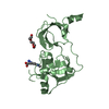
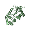
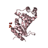
| Deposited unit | 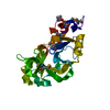
| ||||||||
|---|---|---|---|---|---|---|---|---|---|
| 1 |
| ||||||||
| Unit cell |
|
-Components
| #1: Protein | Mass: 22931.738 Da / Num. of mol.: 1 Source method: isolated from a genetically manipulated source Source: (gene. exp.) Corynebacterium diphtheriae (bacteria) / Plasmid: pET21 / Production host: |
|---|---|
| #2: Water | ChemComp-HOH /  Mass: 18.015 Da / Num. of mol.: 154 / Source method: isolated from a natural source / Formula: H2O Mass: 18.015 Da / Num. of mol.: 154 / Source method: isolated from a natural source / Formula: H2O |
-Experimental details
-Experiment
| Experiment | Method: X-RAY DIFFRACTION / Number of used crystals: 1 |
|---|
- Sample preparation
| Crystal | Density Matthews: 2.13 Å3/Da / Density % sol: 42.26 % Description: THE STRUCTURE FACTOR FILE CONTAINS FRIEDEL PAIRS |
|---|---|
| Crystal grow | Temperature: 293 K / Method: vapor diffusion, hanging drop Details: 4% PEG3350, 200 mM MgCl2, VAPOR DIFFUSION, HANGING DROP, temperature 293K |
-Data collection
| Diffraction | Mean temperature: 100 K |
|---|---|
| Diffraction source | Source: SYNCHROTRON / Site: NSLS  / Beamline: X4A / Wavelength: 0.979 Å / Beamline: X4A / Wavelength: 0.979 Å |
| Detector | Type: ADSC QUANTUM 4 / Detector: CCD / Date: Jul 13, 2006 / Details: mirrors |
| Radiation | Protocol: SINGLE WAVELENGTH / Monochromatic (M) / Laue (L): M / Scattering type: x-ray |
| Radiation wavelength | Wavelength: 0.979 Å / Relative weight: 1 |
| Reflection | Resolution: 1.9→40 Å / Num. obs: 28205 / % possible obs: 93.5 % / Observed criterion σ(I): -3 / Redundancy: 4.1 % / Biso Wilson estimate: 16.7 Å2 / Rmerge(I) obs: 0.088 / Net I/σ(I): 19.8 |
| Reflection shell | Resolution: 1.9→1.97 Å / % possible all: 86 |
- Processing
| Software |
| ||||||||||||||||||||
|---|---|---|---|---|---|---|---|---|---|---|---|---|---|---|---|---|---|---|---|---|---|
| Refinement | Method to determine structure: SAD / Resolution: 1.9→18.76 Å / Rfactor Rfree error: 0.006 / Data cutoff high absF: 123120.9 / Data cutoff low absF: 0 / Isotropic thermal model: RESTRAINED / Cross valid method: THROUGHOUT / σ(F): 2 / Stereochemistry target values: Engh & Huber / Details: The Friedel pairs were used for phasing
| ||||||||||||||||||||
| Solvent computation | Solvent model: FLAT MODEL / Bsol: 47.6486 Å2 / ksol: 0.384251 e/Å3 | ||||||||||||||||||||
| Displacement parameters | Biso mean: 18.5 Å2
| ||||||||||||||||||||
| Refine analyze |
| ||||||||||||||||||||
| Refinement step | Cycle: LAST / Resolution: 1.9→18.76 Å
| ||||||||||||||||||||
| Refine LS restraints |
| ||||||||||||||||||||
| LS refinement shell | Resolution: 1.9→2.02 Å / Rfactor Rfree error: 0.019 / Total num. of bins used: 6
| ||||||||||||||||||||
| Xplor file |
|
