 Movie
Movie Controller

[English] 日本語
 Yorodumi
- PDB-2hql: Crystal structure of a small single-stranded DNA binding protein ... -
+ Open data

- Basic information
| Entry | Database: PDB / ID: 2hql | ||||||
|---|---|---|---|---|---|---|---|
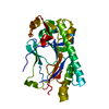
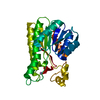
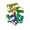
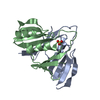
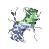
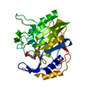
| Title | Crystal structure of a small single-stranded DNA binding protein from Mycoplasma pneumoniae | ||||||
 Components Components | Hypothetical protein MG376 homolog | ||||||
 Keywords Keywords | DNA BINDING PROTEIN / Structural genomics / conserved hypothetical protein / Mycoplasma pneumoniae / GI:1673959 / MPN554 / MG376 / OB fold / Single-stranded DNA binding protein / PSI / Protein Structure Initiative / Berkeley Structural Genomics Center / BSGC | ||||||
| Function / homology | Protein of unknown function DUF3217 / Protein of unknown function (DUF3217) / Nucleic acid-binding proteins / OB fold (Dihydrolipoamide Acetyltransferase, E2P) / Nucleic acid-binding, OB-fold / Beta Barrel / Mainly Beta / Uncharacterized protein MG376 homolog Function and homology information Function and homology information | ||||||
| Biological species |  Mycoplasma pneumoniae (Filterable agent of primary atypical pneumonia) Mycoplasma pneumoniae (Filterable agent of primary atypical pneumonia) | ||||||
| Method |  X-RAY DIFFRACTION / SYNCHROTRON / SAD / Resolution: 2 Å X-RAY DIFFRACTION / SYNCHROTRON / SAD / Resolution: 2 Å | ||||||
 Authors Authors | Das, D. / Berkeley Structural Genomics Center (BSGC) | ||||||
 Citation Citation | Journal: Proteins / Year: 2007 Title: Crystal structure of a novel single-stranded DNA binding protein from Mycoplasma pneumoniae. Authors: Das, D. / Hyun, H. / Lou, Y. / Yokota, H. / Kim, R. / Kim, S.H. | ||||||
| History |
|
- Structure visualization
| Structure viewer | Molecule: MolmilJmol/JSmol |
|---|
- Downloads & links
-Download
| PDBx/mmCIF format | 2hql.cif.gz | 135.4 KB | Display | PDBx/mmCIF format |
|---|---|---|---|---|
| PDB format | pdb2hql.ent.gz | 107.8 KB | Display | PDB format |
| PDBx/mmJSON format | 2hql.json.gz | Tree view | PDBx/mmJSON format | |
| Others |  Other downloads Other downloads |
-Validation report
| Arichive directory | https://data.pdbj.org/pub/pdb/validation_reports/hq/2hqlftp://data.pdbj.org/pub/pdb/validation_reports/hq/2hql | HTTPS FTP |
|---|
-Related structure data
| Similar structure data | |
|---|---|
| Other databases |


-Links
 PDBj
PDBj- Assembly
| Deposited unit | 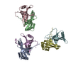
| ||||||||
|---|---|---|---|---|---|---|---|---|---|
| 1 | 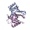
| ||||||||
| 2 | 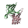
| ||||||||
| 3 | 
| ||||||||
| Unit cell |
|
-Components
| #1: Protein | Mass: 12829.871 Da / Num. of mol.: 6 Source method: isolated from a genetically manipulated source Source: (gene. exp.) Mycoplasma pneumoniae (Filterable agent of primary atypical pneumonia)Gene: MPN554, MP288 / Plasmid: pET21a derivative / Production host: #2: Water | ChemComp-HOH / |  Mass: 18.015 Da / Num. of mol.: 168 / Source method: isolated from a natural source / Formula: H2O Mass: 18.015 Da / Num. of mol.: 168 / Source method: isolated from a natural source / Formula: H2O |
|---|
-Experimental details
-Experiment
| Experiment | Method: X-RAY DIFFRACTION / Number of used crystals: 3 |
|---|
- Sample preparation
| Crystal | Density Matthews: 2.62 Å3/Da / Density % sol: 53 % |
|---|---|
| Crystal grow | Temperature: 298 K / pH: 5.5 Details: 0.2M Ammonium Sulfate, 0.1M Bis-Tris pH 5.5, 25% w/v PEG 3350 (Index Screen 66), VAPOR DIFFUSION, HANGING DROP, temperature 298K, pH 5.50 |
-Data collection
| Diffraction | Mean temperature: 108 K |
|---|---|
| Diffraction source | Source: SYNCHROTRON / Site: ALS  / Beamline: 5.0.2 / Wavelength: 0.99186 / Beamline: 5.0.2 / Wavelength: 0.99186 |
| Detector | Type: ADSC QUANTUM 315 / Detector: CCD / Date: Dec 23, 2004 / Details: MIRRORS |
| Radiation | Protocol: SINGLE WAVELENGTH / Monochromatic (M) / Laue (L): M / Scattering type: x-ray |
| Radiation wavelength | Wavelength: 0.99186 Å / Relative weight: 1 |
| Reflection | Resolution: 2→50 Å / Num. obs: 51433 / % possible obs: 97.7 % / Observed criterion σ(I): -3 / Redundancy: 2.4 % / Biso Wilson estimate: 15 Å2 / Rmerge(I) obs: 0.055 / Rsym value: 0.064 / Net I/σ(I): 13.9 |
| Reflection shell | Resolution: 2→2.07 Å / Redundancy: 2 % / Rmerge(I) obs: 0.304 / Mean I/σ(I) obs: 3.2 / Rsym value: 0.304 / % possible all: 87.7 |
- Processing
| Software |
| ||||||||||||||||||||||||||||||||||||||||||||||||||||||||||||
|---|---|---|---|---|---|---|---|---|---|---|---|---|---|---|---|---|---|---|---|---|---|---|---|---|---|---|---|---|---|---|---|---|---|---|---|---|---|---|---|---|---|---|---|---|---|---|---|---|---|---|---|---|---|---|---|---|---|---|---|---|---|
| Refinement | Method to determine structure: SAD / Resolution: 2→50 Å / σ(F): 2 / Stereochemistry target values: ENGH & HUBER
| ||||||||||||||||||||||||||||||||||||||||||||||||||||||||||||
| Displacement parameters | Biso mean: 31.3 Å2
| ||||||||||||||||||||||||||||||||||||||||||||||||||||||||||||
| Refine analyze |
| ||||||||||||||||||||||||||||||||||||||||||||||||||||||||||||
| Refinement step | Cycle: LAST / Resolution: 2→50 Å
| ||||||||||||||||||||||||||||||||||||||||||||||||||||||||||||
| Refine LS restraints |
| ||||||||||||||||||||||||||||||||||||||||||||||||||||||||||||
| LS refinement shell | Resolution: 2→2.13 Å / Total num. of bins used: 6 |
