 Movie
Movie Controller

[English] 日本語
 Yorodumi
- PDB-2hl8: SUMO protease Ulp1 with the catalytic cysteine oxidized to a sulf... -
+ Open data

- Basic information
| Entry | Database: PDB / ID: 2hl8 | ||||||
|---|---|---|---|---|---|---|---|
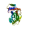
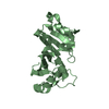
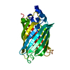
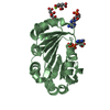
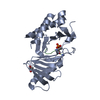
| Title | SUMO protease Ulp1 with the catalytic cysteine oxidized to a sulfinic acid | ||||||
 Components Components | Ubiquitin-like-specific protease 1 | ||||||
 Keywords Keywords | HYDROLASE | ||||||
| Function / homology |  Function and homology information Function and homology informationUlp1 peptidase / deSUMOylase activity / protein desumoylation / SUMO is proteolytically processed / Major pathway of rRNA processing in the nucleolus and cytosol / nuclear pore / cysteine-type peptidase activity / G2/M transition of mitotic cell cycle / nuclear envelope / nucleolus ...Ulp1 peptidase / deSUMOylase activity / protein desumoylation / SUMO is proteolytically processed / Major pathway of rRNA processing in the nucleolus and cytosol / nuclear pore / cysteine-type peptidase activity / G2/M transition of mitotic cell cycle / nuclear envelope / nucleolus / proteolysis / nucleus Similarity search - Function | ||||||
| Biological species |  | ||||||
| Method |  X-RAY DIFFRACTION / MOLECULAR REPLACEMENT / Resolution: 2 Å X-RAY DIFFRACTION / MOLECULAR REPLACEMENT / Resolution: 2 Å | ||||||
 Authors Authors | Xu, Z. / Ng, T.B. / Au, S.W.N. | ||||||
 Citation Citation | Journal: Faseb J. / Year: 2008 Title: Molecular basis of the redox regulation of SUMO proteases: a protective mechanism of intermolecular disulfide linkage against irreversible sulfhydryl oxidation Authors: Xu, Z. / Lam, L.S.M. / Lam, L.H. / Chau, S.F. / Ng, T.B. / Au, S.W.N. #1: Journal: Mol.Cell / Year: 2000Title: Ulp1-SUMO crystal structure and genetic analysis reveal conserved interactions and a regulatory element essential for cell growth in yeast Authors: Mossessova, E. / Lima, C.D. | ||||||
| History |
|
- Structure visualization
| Structure viewer | Molecule: MolmilJmol/JSmol |
|---|
- Downloads & links
-Download
| PDBx/mmCIF format | 2hl8.cif.gz | 59.8 KB | Display | PDBx/mmCIF format |
|---|---|---|---|---|
| PDB format | pdb2hl8.ent.gz | 43.3 KB | Display | PDB format |
| PDBx/mmJSON format | 2hl8.json.gz | Tree view | PDBx/mmJSON format | |
| Others |  Other downloads Other downloads |
-Validation report
| Arichive directory | https://data.pdbj.org/pub/pdb/validation_reports/hl/2hl8ftp://data.pdbj.org/pub/pdb/validation_reports/hl/2hl8 | HTTPS FTP |
|---|
-Related structure data
| Related structure data | 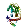 2hkpC  2hl9C  1euvS C: citing same article ( S: Starting model for refinement |
|---|---|
| Similar structure data |


-Links
 PDBj
PDBj

- Assembly
| Deposited unit | 
| ||||||||
|---|---|---|---|---|---|---|---|---|---|
| 1 |
| ||||||||
| Unit cell |
|
-Components
| #1: Protein | Mass: 25682.299 Da / Num. of mol.: 1 / Fragment: c-terminal catalytic domain Source method: isolated from a genetically manipulated source Source: (gene. exp.) Plasmid: pGEX-6P-2 / Production host:  References: UniProt: Q02724, Hydrolases; Acting on peptide bonds (peptidases); Cysteine endopeptidases |
|---|---|
| #2: Water | ChemComp-HOH /  Mass: 18.015 Da / Num. of mol.: 116 / Source method: isolated from a natural source / Formula: H2O Mass: 18.015 Da / Num. of mol.: 116 / Source method: isolated from a natural source / Formula: H2O |
| Has protein modification | Y |
-Experimental details
-Experiment
| Experiment | Method: X-RAY DIFFRACTION / Number of used crystals: 1 |
|---|
- Sample preparation
| Crystal | Density Matthews: 2.11 Å3/Da / Density % sol: 41.62 % |
|---|---|
| Crystal grow | Temperature: 289 K / Method: vapor diffusion, hanging drop / pH: 6.5 Details: 20%(w/v) PEG 3350, 0.2M NaCl, pH 6.5, VAPOR DIFFUSION, HANGING DROP, temperature 289K |
-Data collection
| Diffraction | Mean temperature: 100 K |
|---|---|
| Diffraction source | Source: ROTATING ANODE / Type: RIGAKU / Wavelength: 1.54 Å |
| Detector | Type: RIGAKU RAXIS IV / Detector: IMAGE PLATE / Date: Mar 28, 2006 / Details: mirrors |
| Radiation | Monochromator: VariMax HR / Protocol: SINGLE WAVELENGTH / Monochromatic (M) / Laue (L): M / Scattering type: x-ray |
| Radiation wavelength | Wavelength: 1.54 Å / Relative weight: 1 |
| Reflection | Resolution: 2→44.63 Å / Num. obs: 14290 / % possible obs: 97.2 % / Redundancy: 7.35 % / Rmerge(I) obs: 0.073 / Χ2: 0.99 / Net I/σ(I): 16.3 / Scaling rejects: 794 |
| Reflection shell | Resolution: 2→2.07 Å / Redundancy: 7.28 % / Rmerge(I) obs: 0.359 / Mean I/σ(I) obs: 5.1 / Num. measured all: 10146 / Num. unique all: 1393 / Χ2: 1.01 / % possible all: 95.5 |
- Processing
| Software |
| |||||||||||||||||||||||||||||||||
|---|---|---|---|---|---|---|---|---|---|---|---|---|---|---|---|---|---|---|---|---|---|---|---|---|---|---|---|---|---|---|---|---|---|---|
| Refinement | Method to determine structure: MOLECULAR REPLACEMENT Starting model: PDB ENTRY 1EUV Resolution: 2→19.83 Å / Cor.coef. Fo:Fc: 0.952 / Cor.coef. Fo:Fc free: 0.936 / SU B: 4.897 / SU ML: 0.136 / Cross valid method: THROUGHOUT / σ(F): 0 / ESU R: 0.221 / ESU R Free: 0.179 Stereochemistry target values: MAXIMUM LIKELIHOOD WITH PHASES Details: HYDROGENS HAVE BEEN ADDED IN THE RIDING POSITIONS
| |||||||||||||||||||||||||||||||||
| Solvent computation | Ion probe radii: 0.8 Å / Shrinkage radii: 0.8 Å / VDW probe radii: 1.2 Å / Solvent model: MASK | |||||||||||||||||||||||||||||||||
| Displacement parameters | Biso mean: 29.135 Å2
| |||||||||||||||||||||||||||||||||
| Refinement step | Cycle: LAST / Resolution: 2→19.83 Å
| |||||||||||||||||||||||||||||||||
| Refine LS restraints |
| |||||||||||||||||||||||||||||||||
| LS refinement shell | Resolution: 2→2.052 Å / Total num. of bins used: 20
|
