 Movie
Movie Controller

[English] 日本語
 Yorodumi
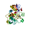
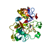
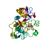
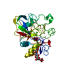
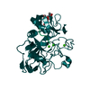
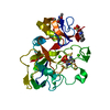
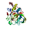
- PDB-2hib: human formylglycine generating enzyme, C336S mutant, iodide co-cr... -
+ Open data

- Basic information
| Entry | Database: PDB / ID: 2hib | |||||||||
|---|---|---|---|---|---|---|---|---|---|---|
| Title | human formylglycine generating enzyme, C336S mutant, iodide co-crystallization | |||||||||
 Components Components | Sulfatase-modifying factor 1 | |||||||||
 Keywords Keywords | HYDROLASE ACTIVATOR / PROTEIN BINDING / formylglycine / post-translational modification / endoplasmic reticulum / sulfatase | |||||||||
| Function / homology |  Function and homology information Function and homology informationThe activation of arylsulfatases / formylglycine-generating enzyme / formylglycine-generating oxidase activity / protein oxidation / glycosphingolipid catabolic process / Glycosphingolipid catabolism / cupric ion binding / post-translational protein modification / oxidoreductase activity / endoplasmic reticulum lumen ...The activation of arylsulfatases / formylglycine-generating enzyme / formylglycine-generating oxidase activity / protein oxidation / glycosphingolipid catabolic process / Glycosphingolipid catabolism / cupric ion binding / post-translational protein modification / oxidoreductase activity / endoplasmic reticulum lumen / endoplasmic reticulum / membrane / identical protein binding Similarity search - Function | |||||||||
| Biological species |  Homo sapiens (human) Homo sapiens (human) | |||||||||
| Method |  X-RAY DIFFRACTION / MOLECULAR REPLACEMENT / Resolution: 2 Å X-RAY DIFFRACTION / MOLECULAR REPLACEMENT / Resolution: 2 Å | |||||||||
 Authors Authors | Rudolph, M.G. / Roeser, D. | |||||||||
 Citation Citation | Journal: Acta Crystallogr.,Sect.D / Year: 2007 Title: Probing the oxygen-binding site of the human formylglycine-generating enzyme using halide ions. Authors: Roeser, D. / Schmidt, B. / Preusser-Kunze, A. / Rudolph, M.G. | |||||||||
| History |
|
- Structure visualization
| Structure viewer | Molecule: MolmilJmol/JSmol |
|---|
- Downloads & links
-Download
| PDBx/mmCIF format | 2hib.cif.gz | 77.2 KB | Display | PDBx/mmCIF format |
|---|---|---|---|---|
| PDB format | pdb2hib.ent.gz | 55.6 KB | Display | PDB format |
| PDBx/mmJSON format | 2hib.json.gz | Tree view | PDBx/mmJSON format | |
| Others |  Other downloads Other downloads |
-Validation report
| Arichive directory | https://data.pdbj.org/pub/pdb/validation_reports/hi/2hibftp://data.pdbj.org/pub/pdb/validation_reports/hi/2hib | HTTPS FTP |
|---|
-Related structure data
| Related structure data |  2hi8C  2aijS S: Starting model for refinement C: citing same article ( |
|---|---|
| Similar structure data |


-Links
 PDBj
PDBj


- Assembly
| Deposited unit | 
| ||||||||
|---|---|---|---|---|---|---|---|---|---|
| 1 |
| ||||||||
| Unit cell |
| ||||||||
| Components on special symmetry positions |
|
-Components
| #1: Protein | Mass: 32110.613 Da / Num. of mol.: 1 / Fragment: RESIDUES 86-371 / Mutation: C336S Source method: isolated from a genetically manipulated source Source: (gene. exp.) Homo sapiens (human) / Cell (production host): fibrosarcoma cells / Production host: Homo sapiens (human) / References: UniProt: Q8NBK3 | ||||||
|---|---|---|---|---|---|---|---|
| #2: Polysaccharide | 2-acetamido-2-deoxy-beta-D-glucopyranose-(1-4)-2-acetamido-2-deoxy-beta-D-glucopyranose Source method: isolated from a genetically manipulated source | ||||||
| #3: Chemical |   Mass: 40.078 Da / Num. of mol.: 2 / Source method: obtained synthetically / Formula: Ca Mass: 40.078 Da / Num. of mol.: 2 / Source method: obtained synthetically / Formula: Ca#4: Chemical | ChemComp-IOD /   Mass: 126.904 Da / Num. of mol.: 11 / Source method: obtained synthetically / Formula: I Mass: 126.904 Da / Num. of mol.: 11 / Source method: obtained synthetically / Formula: I#5: Water | ChemComp-HOH / |  Mass: 18.015 Da / Num. of mol.: 258 / Source method: isolated from a natural source / Formula: H2O Mass: 18.015 Da / Num. of mol.: 258 / Source method: isolated from a natural source / Formula: H2OHas protein modification | Y | |
-Experimental details
-Experiment
| Experiment | Method: X-RAY DIFFRACTION / Number of used crystals: 2 |
|---|
- Sample preparation
| Crystal | Density Matthews: 2.28 Å3/Da / Density % sol: 46.17 % |
|---|---|
| Crystal grow | Temperature: 293 K / Method: vapor diffusion, sitting drop / pH: 8.5 Details: PEG 4000, Calcium iodide, TRIS, pH 8.5, VAPOR DIFFUSION, SITTING DROP, temperature 293K |
-Data collection
| Diffraction | Mean temperature: 100 K |
|---|---|
| Diffraction source | Source: ROTATING ANODE / Type: RIGAKU / Wavelength: 1.5419 Å |
| Detector | Type: MAR scanner 345 mm plate / Detector: IMAGE PLATE / Date: May 15, 2006 / Details: osmic mirrors |
| Radiation | Protocol: SINGLE WAVELENGTH / Monochromatic (M) / Laue (L): M / Scattering type: x-ray |
| Radiation wavelength | Wavelength: 1.5419 Å / Relative weight: 1 |
| Reflection | Resolution: 1.99→50 Å / Num. obs: 72271 / % possible obs: 96.8 % / Observed criterion σ(F): 0 / Observed criterion σ(I): 0 / Redundancy: 3.6 % / Rmerge(I) obs: 0.132 / Net I/σ(I): 9 |
| Reflection shell | Resolution: 1.99→2.06 Å / Redundancy: 2.3 % / Rmerge(I) obs: 0.413 / Mean I/σ(I) obs: 1.9 / Num. unique all: 1570 |
- Processing
| Software |
| ||||||||||||||||||||||||||||||||||||||||||||||||||||||||||||||||||||||||||||||||||||||||||||||||||||||||||||||||||||||||||||||||||
|---|---|---|---|---|---|---|---|---|---|---|---|---|---|---|---|---|---|---|---|---|---|---|---|---|---|---|---|---|---|---|---|---|---|---|---|---|---|---|---|---|---|---|---|---|---|---|---|---|---|---|---|---|---|---|---|---|---|---|---|---|---|---|---|---|---|---|---|---|---|---|---|---|---|---|---|---|---|---|---|---|---|---|---|---|---|---|---|---|---|---|---|---|---|---|---|---|---|---|---|---|---|---|---|---|---|---|---|---|---|---|---|---|---|---|---|---|---|---|---|---|---|---|---|---|---|---|---|---|---|---|---|
| Refinement | Method to determine structure: MOLECULAR REPLACEMENT Starting model: 2aij Resolution: 2→33.83 Å / Cor.coef. Fo:Fc: 0.951 / Cor.coef. Fo:Fc free: 0.91 / SU B: 4.181 / SU ML: 0.118 / Cross valid method: THROUGHOUT / ESU R: 0.185 / ESU R Free: 0.176 / Stereochemistry target values: MAXIMUM LIKELIHOOD Details: HYDROGENS HAVE BEEN ADDED IN THE RIDING POSITIONS. The close contacts are partial occupancy contacts, where the occupancies of the clashing atoms sum up to one.
| ||||||||||||||||||||||||||||||||||||||||||||||||||||||||||||||||||||||||||||||||||||||||||||||||||||||||||||||||||||||||||||||||||
| Solvent computation | Ion probe radii: 0.8 Å / Shrinkage radii: 0.8 Å / VDW probe radii: 1.2 Å / Solvent model: BABINET MODEL WITH MASK | ||||||||||||||||||||||||||||||||||||||||||||||||||||||||||||||||||||||||||||||||||||||||||||||||||||||||||||||||||||||||||||||||||
| Displacement parameters | Biso mean: 25.822 Å2
| ||||||||||||||||||||||||||||||||||||||||||||||||||||||||||||||||||||||||||||||||||||||||||||||||||||||||||||||||||||||||||||||||||
| Refinement step | Cycle: LAST / Resolution: 2→33.83 Å
| ||||||||||||||||||||||||||||||||||||||||||||||||||||||||||||||||||||||||||||||||||||||||||||||||||||||||||||||||||||||||||||||||||
| Refine LS restraints |
| ||||||||||||||||||||||||||||||||||||||||||||||||||||||||||||||||||||||||||||||||||||||||||||||||||||||||||||||||||||||||||||||||||
| LS refinement shell | Resolution: 2→2.052 Å / Total num. of bins used: 20
|
