 Movie
Movie Controller

[English] 日本語
 Yorodumi
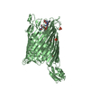
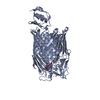
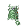
- PDB-2hdi: Crystal structure of the Colicin I receptor Cir from E.coli in co... -
+ Open data

- Basic information
| Entry | Database: PDB / ID: 2hdi | ||||||
|---|---|---|---|---|---|---|---|
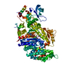
| Title | Crystal structure of the Colicin I receptor Cir from E.coli in complex with receptor binding domain of Colicin Ia. | ||||||
 Components Components |
| ||||||
 Keywords Keywords | Protein Transport / Antimicrobial Protein / Outer membrane / Iron transport / TonB box / Signal transduction / Colicin I Receptor / Receptor Ligand / Membrane Protein | ||||||
| Function / homology |  Function and homology information Function and homology informationsiderophore-iron transmembrane transporter activity / colicin transmembrane transporter activity / pore-forming activity / siderophore transmembrane transport / siderophore uptake transmembrane transporter activity / transmembrane transporter complex / cell outer membrane / killing of cells of another organism / defense response to Gram-negative bacterium / intracellular iron ion homeostasis ...siderophore-iron transmembrane transporter activity / colicin transmembrane transporter activity / pore-forming activity / siderophore transmembrane transport / siderophore uptake transmembrane transporter activity / transmembrane transporter complex / cell outer membrane / killing of cells of another organism / defense response to Gram-negative bacterium / intracellular iron ion homeostasis / membrane / plasma membrane Similarity search - Function | ||||||
| Biological species |  | ||||||
| Method |  X-RAY DIFFRACTION / SYNCHROTRON / MOLECULAR REPLACEMENT / Resolution: 2.5 Å X-RAY DIFFRACTION / SYNCHROTRON / MOLECULAR REPLACEMENT / Resolution: 2.5 Å | ||||||
 Authors Authors | Buchanan, S.K. / Esser, L. / Lukacik, P. | ||||||
 Citation Citation | Journal: Embo J. / Year: 2007 Title: Structure of colicin I receptor bound to the R-domain of colicin Ia: implications for protein import. Authors: Buchanan, S.K. / Lukacik, P. / Grizot, S. / Ghirlando, R. / Ali, M.M. / Barnard, T.J. / Jakes, K.S. / Kienker, P.K. / Esser, L. | ||||||
| History |
|
- Structure visualization
| Structure viewer | Molecule: MolmilJmol/JSmol |
|---|
- Downloads & links
-Download
| PDBx/mmCIF format | 2hdi.cif.gz | 160.9 KB | Display | PDBx/mmCIF format |
|---|---|---|---|---|
| PDB format | pdb2hdi.ent.gz | 123.5 KB | Display | PDB format |
| PDBx/mmJSON format | 2hdi.json.gz | Tree view | PDBx/mmJSON format | |
| Others |  Other downloads Other downloads |
-Validation report
| Arichive directory | https://data.pdbj.org/pub/pdb/validation_reports/hd/2hdiftp://data.pdbj.org/pub/pdb/validation_reports/hd/2hdi | HTTPS FTP |
|---|
-Related structure data
| Related structure data | 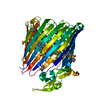 2hdfSC S: Starting model for refinement C: citing same article ( |
|---|---|
| Similar structure data |

-Links
 PDBj
PDBj- Assembly
| Deposited unit | 
| ||||||||
|---|---|---|---|---|---|---|---|---|---|
| 1 |
| ||||||||
| Unit cell |
|
-Components
| #1: Protein | Mass: 71294.242 Da / Num. of mol.: 1 / Fragment: Colicin I receptor / Mutation: W338M, L343M, F589M, V591M Source method: isolated from a genetically manipulated source Source: (gene. exp.) | ||||
|---|---|---|---|---|---|
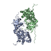
| #2: Protein | Mass: 12891.374 Da / Num. of mol.: 1 / Fragment: R domain of Colicin Ia Source method: isolated from a genetically manipulated source Source: (gene. exp.) | ||||
| #3: Chemical |   Mass: 229.402 Da / Num. of mol.: 2 / Source method: obtained synthetically / Formula: C14H31NO / Comment: LDAO, detergent*YM Mass: 229.402 Da / Num. of mol.: 2 / Source method: obtained synthetically / Formula: C14H31NO / Comment: LDAO, detergent*YM#4: Water | ChemComp-HOH / |  Mass: 18.015 Da / Num. of mol.: 255 / Source method: isolated from a natural source / Formula: H2O Mass: 18.015 Da / Num. of mol.: 255 / Source method: isolated from a natural source / Formula: H2OHas protein modification | Y | |
-Experimental details
-Experiment
| Experiment | Method: X-RAY DIFFRACTION / Number of used crystals: 1 |
|---|
- Sample preparation
| Crystal | Density Matthews: 2.84 Å3/Da / Density % sol: 56.62 % |
|---|---|
| Crystal grow | Temperature: 294 K / Method: vapor diffusion, hanging drop / pH: 6.2 Details: 9 G/L PROTEIN IN 0.02M TRIS PH7.5, 0.2M NACL, 0.05% LDAO, 0.45% C8E4, 3% HEPTANETRIOL MIXED AT 1:1 RATIO WITH 0.1M MES PH6.2, 10% GLYCEROL, 22% PEG 2000 MME, VAPOR DIFFUSION, HANGING DROP, temperature 294K |
-Data collection
| Diffraction | Mean temperature: 100 K |
|---|---|
| Diffraction source | Source: SYNCHROTRON / Site: APS  / Beamline: 22-ID / Wavelength: 1 Å / Beamline: 22-ID / Wavelength: 1 Å |
| Detector | Type: MARMOSAIC 300 mm CCD / Detector: CCD / Date: Oct 24, 2004 |
| Radiation | Monochromator: Si 220 (Rosenbaum-Rock double-crystal monochromator) Protocol: SINGLE WAVELENGTH / Monochromatic (M) / Laue (L): M / Scattering type: x-ray |
| Radiation wavelength | Wavelength: 1 Å / Relative weight: 1 |
| Reflection | Resolution: 2.5→30 Å / Num. all: 32311 / Num. obs: 32311 / % possible obs: 99.9 % / Observed criterion σ(F): 0 / Observed criterion σ(I): 0 / Redundancy: 4.1 % / Rmerge(I) obs: 0.103 / Net I/σ(I): 15.3 |
| Reflection shell | Resolution: 2.5→2.59 Å / Redundancy: 4 % / Rmerge(I) obs: 0.57 / Mean I/σ(I) obs: 2.73 / Num. unique all: 3632 / % possible all: 99.9 |
- Processing
| Software |
| ||||||||||||||||||||||||||||||||||||||||||||||||||||||||||||||||||||||||||||||||||||||||||||||||||||
|---|---|---|---|---|---|---|---|---|---|---|---|---|---|---|---|---|---|---|---|---|---|---|---|---|---|---|---|---|---|---|---|---|---|---|---|---|---|---|---|---|---|---|---|---|---|---|---|---|---|---|---|---|---|---|---|---|---|---|---|---|---|---|---|---|---|---|---|---|---|---|---|---|---|---|---|---|---|---|---|---|---|---|---|---|---|---|---|---|---|---|---|---|---|---|---|---|---|---|---|---|---|
| Refinement | Method to determine structure: MOLECULAR REPLACEMENT Starting model: 2HDF Resolution: 2.5→15 Å / Cor.coef. Fo:Fc: 0.943 / Cor.coef. Fo:Fc free: 0.897 / SU B: 9.389 / SU ML: 0.112 / TLS residual ADP flag: LIKELY RESIDUAL / Cross valid method: THROUGHOUT / σ(F): 0 / σ(I): 0 / ESU R: 0.48 / ESU R Free: 0.287 / Stereochemistry target values: MAXIMUM LIKELIHOOD / Details: HYDROGENS HAVE BEEN ADDED IN THE RIDING POSITIONS
| ||||||||||||||||||||||||||||||||||||||||||||||||||||||||||||||||||||||||||||||||||||||||||||||||||||
| Solvent computation | Ion probe radii: 0.8 Å / Shrinkage radii: 0.8 Å / VDW probe radii: 1.2 Å / Solvent model: BABINET MODEL WITH MASK | ||||||||||||||||||||||||||||||||||||||||||||||||||||||||||||||||||||||||||||||||||||||||||||||||||||
| Displacement parameters | Biso mean: 44.618 Å2
| ||||||||||||||||||||||||||||||||||||||||||||||||||||||||||||||||||||||||||||||||||||||||||||||||||||
| Refinement step | Cycle: LAST / Resolution: 2.5→15 Å
| ||||||||||||||||||||||||||||||||||||||||||||||||||||||||||||||||||||||||||||||||||||||||||||||||||||
| Refine LS restraints |
| ||||||||||||||||||||||||||||||||||||||||||||||||||||||||||||||||||||||||||||||||||||||||||||||||||||
| LS refinement shell | Resolution: 2.5→2.563 Å / Total num. of bins used: 20
| ||||||||||||||||||||||||||||||||||||||||||||||||||||||||||||||||||||||||||||||||||||||||||||||||||||
| Refinement TLS params. | Method: refined / Refine-ID: X-RAY DIFFRACTION
| ||||||||||||||||||||||||||||||||||||||||||||||||||||||||||||||||||||||||||||||||||||||||||||||||||||
| Refinement TLS group |
|
