 Movie
Movie Controller

[English] 日本語
 Yorodumi
- PDB-2h1n: 3.0 A X-ray structure of putative oligoendopeptidase F: crystals ... -
+ Open data

- Basic information
| Entry | Database: PDB / ID: 2h1n | ||||||
|---|---|---|---|---|---|---|---|
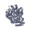
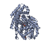
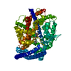
| Title | 3.0 A X-ray structure of putative oligoendopeptidase F: crystals grown by vapor diffusion technique | ||||||
 Components Components | Oligoendopeptidase F | ||||||
 Keywords Keywords | HYDROLASE / STRUCTURAL GENOMICS / PROTEIN STRUCTURE INITIATIVE / MIDWEST CENTER FOR STRUCTURAL GENOMICS / MCSG / PSI | ||||||
| Function / homology |  Function and homology information Function and homology informationpeptide metabolic process / Hydrolases; Acting on peptide bonds (peptidases); Metalloendopeptidases / metalloendopeptidase activity / proteolysis / metal ion binding Similarity search - Function | ||||||
| Biological species |   Geobacillus stearothermophilus (bacteria) Geobacillus stearothermophilus (bacteria) | ||||||
| Method |  X-RAY DIFFRACTION / SYNCHROTRON / SAD / Resolution: 3 Å X-RAY DIFFRACTION / SYNCHROTRON / SAD / Resolution: 3 Å | ||||||
 Authors Authors | Gerdts, C.J. / Tereshko, V. / Dementieva, I. / Collart, F. / Joachimiak, A. / Kossiakoff, A. / Ismagilov, R.F. / Midwest Center for Structural Genomics (MCSG) | ||||||
 Citation Citation | Journal: Angew.Chem.Int.Ed.Engl. / Year: 2006 Title: Time-Controlled Microfluidic Seeding in nL-Volume Droplets To Separate Nucleation and Growth Stages of Protein Crystallization. Authors: Gerdts, C.J. / Tereshko, V. / Yadav, M.K. / Dementieva, I. / Collart, F. / Joachimiak, A. / Stevens, R.C. / Kuhn, P. / Kossiakoff, A. / Ismagilov, R.F. | ||||||
| History |
| ||||||
| Remark 999 | SEQUENCE At the time of processing, the sequence of this protein is not available at the UNP ...SEQUENCE At the time of processing, the sequence of this protein is not available at the UNP sequence database. Residues -2 to 0 are cloning artifacts. | ||||||
| Remark 300 | BIOMOLECULE: 1, 2 THIS ENTRY CONTAINS THE CRYSTALLOGRAPHIC ASYMMETRIC UNIT WHICH CONSISTS OF 2 ...BIOMOLECULE: 1, 2 THIS ENTRY CONTAINS THE CRYSTALLOGRAPHIC ASYMMETRIC UNIT WHICH CONSISTS OF 2 CHAIN(S). EACH CHAIN REPRESENTS ONE BIOLOGICAL UNIT. THE OLIGOMERIZATION IS UNKNOWN. |
- Structure visualization
| Structure viewer | Molecule: MolmilJmol/JSmol |
|---|
- Downloads & links
-Download
| PDBx/mmCIF format | 2h1n.cif.gz | 235.5 KB | Display | PDBx/mmCIF format |
|---|---|---|---|---|
| PDB format | pdb2h1n.ent.gz | 190.9 KB | Display | PDB format |
| PDBx/mmJSON format | 2h1n.json.gz | Tree view | PDBx/mmJSON format | |
| Others |  Other downloads Other downloads |
-Validation report
| Arichive directory | https://data.pdbj.org/pub/pdb/validation_reports/h1/2h1nftp://data.pdbj.org/pub/pdb/validation_reports/h1/2h1n | HTTPS FTP |
|---|
-Related structure data


-Links
 PDBj
PDBj

- Assembly
| Deposited unit | 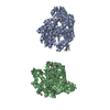
| |||||||||||||||||||||||||||||||||||||||||||||||||||||||||||||||||||||||||||||||||||||||||||||||||||||||||||||||||||||||||||||||||||||||||||||||||||||||||||||||||||||||||||||||||||||||||||||||||||
|---|---|---|---|---|---|---|---|---|---|---|---|---|---|---|---|---|---|---|---|---|---|---|---|---|---|---|---|---|---|---|---|---|---|---|---|---|---|---|---|---|---|---|---|---|---|---|---|---|---|---|---|---|---|---|---|---|---|---|---|---|---|---|---|---|---|---|---|---|---|---|---|---|---|---|---|---|---|---|---|---|---|---|---|---|---|---|---|---|---|---|---|---|---|---|---|---|---|---|---|---|---|---|---|---|---|---|---|---|---|---|---|---|---|---|---|---|---|---|---|---|---|---|---|---|---|---|---|---|---|---|---|---|---|---|---|---|---|---|---|---|---|---|---|---|---|---|---|---|---|---|---|---|---|---|---|---|---|---|---|---|---|---|---|---|---|---|---|---|---|---|---|---|---|---|---|---|---|---|---|---|---|---|---|---|---|---|---|---|---|---|---|---|---|---|---|---|
| 1 | 
| |||||||||||||||||||||||||||||||||||||||||||||||||||||||||||||||||||||||||||||||||||||||||||||||||||||||||||||||||||||||||||||||||||||||||||||||||||||||||||||||||||||||||||||||||||||||||||||||||||
| 2 | 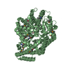
| |||||||||||||||||||||||||||||||||||||||||||||||||||||||||||||||||||||||||||||||||||||||||||||||||||||||||||||||||||||||||||||||||||||||||||||||||||||||||||||||||||||||||||||||||||||||||||||||||||
| Unit cell |
| |||||||||||||||||||||||||||||||||||||||||||||||||||||||||||||||||||||||||||||||||||||||||||||||||||||||||||||||||||||||||||||||||||||||||||||||||||||||||||||||||||||||||||||||||||||||||||||||||||
| Noncrystallographic symmetry (NCS) | NCS domain:
NCS domain segments: Ens-ID: 1 / Refine code: 2
|