regulation of transcriptional start site selection at RNA polymerase II promoter / nucleolar chromatin / negative regulation of DNA-templated DNA replication / regulation of chromatin organization / SLIK (SAGA-like) complex / DNA double-strand break processing / rDNA binding / nucleosome organization / nucleosome array spacer activity / histone H3K4me3 reader activity ...regulation of transcriptional start site selection at RNA polymerase II promoter / nucleolar chromatin / negative regulation of DNA-templated DNA replication / regulation of chromatin organization / SLIK (SAGA-like) complex / DNA double-strand break processing / rDNA binding / nucleosome organization / nucleosome array spacer activity / histone H3K4me3 reader activity / SAGA complex / ATP-dependent chromatin remodeler activity / sister chromatid cohesion / termination of RNA polymerase II transcription / termination of RNA polymerase I transcription / ATP-dependent activity, acting on DNA / transcription elongation by RNA polymerase II / chromatin DNA binding / double-strand break repair via homologous recombination / Hydrolases; Acting on acid anhydrides; Acting on acid anhydrides to facilitate cellular and subcellular movement / site of double-strand break / histone binding / transcription cis-regulatory region binding / chromatin remodeling / chromatin binding / regulation of transcription by RNA polymerase II / chromatin / ATP hydrolysis activity / mitochondrion / DNA binding / ATP binding / nucleus Similarity search - Function
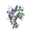
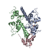
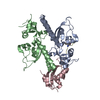
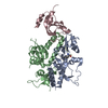
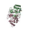
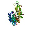
BIOMOLECULE: 1 THIS ENTRY CONTAINS THE CRYSTALLOGRAPHIC ASYMMETRIC UNIT WHICH CONSISTS OF 2 CHAIN(S) ...BIOMOLECULE: 1 THIS ENTRY CONTAINS THE CRYSTALLOGRAPHIC ASYMMETRIC UNIT WHICH CONSISTS OF 2 CHAIN(S). ACCORDING TO AUTHORS, THE BIOLOGICAL UNIT OF THE PROTEIN IN VIVO IS UNKNOWN
In the structure databanks used in Yorodumi, some data are registered as the other names, "COVID-19 virus" and "2019-nCoV". Here are the details of the virus and the list of structure data.
Jan 31, 2019. EMDB accession codes are about to change! (news from PDBe EMDB page)
EMDB accession codes are about to change! (news from PDBe EMDB page)
The allocation of 4 digits for EMDB accession codes will soon come to an end. Whilst these codes will remain in use, new EMDB accession codes will include an additional digit and will expand incrementally as the available range of codes is exhausted. The current 4-digit format prefixed with “EMD-” (i.e. EMD-XXXX) will advance to a 5-digit format (i.e. EMD-XXXXX), and so on. It is currently estimated that the 4-digit codes will be depleted around Spring 2019, at which point the 5-digit format will come into force.
The EM Navigator/Yorodumi systems omit the EMD- prefix.
Related info.:Q: What is EMD? / ID/Accession-code notation in Yorodumi/EM Navigator
Yorodumi is a browser for structure data from EMDB, PDB, SASBDB, etc.
This page is also the successor to EM Navigator detail page, and also detail information page/front-end page for Omokage search.
The word "yorodu" (or yorozu) is an old Japanese word meaning "ten thousand". "mi" (miru) is to see.
Related info.:EMDB / PDB / SASBDB / Comparison of 3 databanks / Yorodumi Search / Aug 31, 2016. New EM Navigator & Yorodumi / Yorodumi Papers / Jmol/JSmol / Function and homology information / Changes in new EM Navigator and Yorodumi
 Movie
Movie Controller


 Open data
 Basic information
 Components
 Keywords
 Function and homology information
 X-RAY DIFFRACTION /
  Authors
 Citation
 Structure visualization
 Downloads & links
 Other downloads
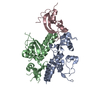


 PDBj
PDBj Assembly


 Mass: 18.015 Da / Num. of mol.: 194 / Source method: isolated from a natural source / Formula: H2O
 Sample preparation
 / Beamline: 22-ID / Wavelength: 1
  Processing