Group: Data collection / Category: chem_comp_atom / chem_comp_bond
Remark 400
COMPOUND RESDIUES A168 AND S169 IN CHAIN A ARE UNIDENTIFIED PART OF C-TERMINUS OF EITHER CHAIN A OR CHAIN B.
Remark 300
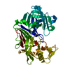
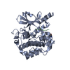
BIOMOLECULE: 1 THIS ENTRY CONTAINS THE CRYSTALLOGRAPHIC ASYMMETRIC UNIT WHICH CONSISTS OF 1 CHAIN. ...BIOMOLECULE: 1 THIS ENTRY CONTAINS THE CRYSTALLOGRAPHIC ASYMMETRIC UNIT WHICH CONSISTS OF 1 CHAIN. THE BIOLOGICAL UNIT OF THIS PROTEIN IS UNKNOWN.
Resolution: 1.4→1.43 Å / Redundancy: 2.2 % / Rmerge(I) obs: 0.513 / Mean I/σ(I) obs: 1.82 / % possible all: 54.3
-
Processing
Software
Name
Version
Classification
REFMAC
5.2.0019
refinement
SBC-Collect
datacollection
HKL-2000
datascaling
SHELXD
phasing
CNS
phasing
ARP/wARP
modelbuilding
Refinement
Method to determine structure: SAD / Resolution: 1.4→32.7 Å / Cor.coef. Fo:Fc: 0.972 / SU B: 1.394 / SU ML: 0.024 / σ(F): 0 / σ(I): 0 / ESU R: 0.06 / Stereochemistry target values: MAXIMUM LIKELIHOOD Details: HYDROGENS HAVE BEEN ADDED IN THE RIDING POSITIONS. ALL DATA WERE USED IN FINAL ROUND OF REFINEMENT. R-FACTOR-ALL CORRESPONDS TO DEPOSITED FILE. R-WORK AND R-FREE FACTORS ARE TAKEN FROM ...Details: HYDROGENS HAVE BEEN ADDED IN THE RIDING POSITIONS. ALL DATA WERE USED IN FINAL ROUND OF REFINEMENT. R-FACTOR-ALL CORRESPONDS TO DEPOSITED FILE. R-WORK AND R-FREE FACTORS ARE TAKEN FROM SECOND TO LAST ROUND OF REFINEMENT WHICH USED TEST DATA SET.
Rfactor
Num. reflection
% reflection
Selection details
Rfree
0.1757
3273
-
RANDOM
Rwork
0.1398
-
-
-
all
0.14
64894
-
-
obs
0.14
64894
92.06 %
-
Solvent computation
Ion probe radii: 0.8 Å / Shrinkage radii: 0.8 Å / VDW probe radii: 1.2 Å / Solvent model: MASK
Displacement parameters
Biso mean: 17.557 Å2
Baniso -1
Baniso -2
Baniso -3
1-
0.05 Å2
0 Å2
0 Å2
2-
-
-0.06 Å2
0 Å2
3-
-
-
0.01 Å2
Refinement step
Cycle: LAST / Resolution: 1.4→32.7 Å
Protein
Nucleic acid
Ligand
Solvent
Total
Num. atoms
2500
0
36
394
2930
Refine LS restraints
Refine-ID
Type
Dev ideal
Dev ideal target
Number
X-RAY DIFFRACTION
r_bond_refined_d
0.017
0.022
2868
X-RAY DIFFRACTION
r_bond_other_d
X-RAY DIFFRACTION
r_angle_refined_deg
1.528
1.97
3934
X-RAY DIFFRACTION
r_angle_other_deg
X-RAY DIFFRACTION
r_dihedral_angle_1_deg
4.402
5
402
X-RAY DIFFRACTION
r_dihedral_angle_2_deg
36.621
26.194
155
X-RAY DIFFRACTION
r_dihedral_angle_3_deg
13.482
15
566
X-RAY DIFFRACTION
r_dihedral_angle_4_deg
16.467
15
15
X-RAY DIFFRACTION
r_chiral_restr
0.104
0.2
449
X-RAY DIFFRACTION
r_gen_planes_refined
0.008
0.02
2224
X-RAY DIFFRACTION
r_gen_planes_other
X-RAY DIFFRACTION
r_nbd_refined
0.237
0.2
1701
X-RAY DIFFRACTION
r_nbd_other
X-RAY DIFFRACTION
r_nbtor_refined
0.309
0.2
1989
X-RAY DIFFRACTION
r_nbtor_other
X-RAY DIFFRACTION
r_xyhbond_nbd_refined
0.312
0.2
362
X-RAY DIFFRACTION
r_xyhbond_nbd_other
X-RAY DIFFRACTION
r_metal_ion_refined
0.185
0.2
22
X-RAY DIFFRACTION
r_metal_ion_other
X-RAY DIFFRACTION
r_symmetry_vdw_refined
0.273
0.2
116
X-RAY DIFFRACTION
r_symmetry_vdw_other
X-RAY DIFFRACTION
r_symmetry_hbond_refined
0.367
0.2
40
X-RAY DIFFRACTION
r_symmetry_hbond_other
X-RAY DIFFRACTION
r_symmetry_metal_ion_refined
0.532
0.2
4
X-RAY DIFFRACTION
r_symmetry_metal_ion_other
X-RAY DIFFRACTION
r_mcbond_it
1.608
1.5
1735
X-RAY DIFFRACTION
r_mcbond_other
X-RAY DIFFRACTION
r_mcangle_it
2.429
2
2829
X-RAY DIFFRACTION
r_scbond_it
3.69
3
1152
X-RAY DIFFRACTION
r_scangle_it
5.418
4.5
1058
X-RAY DIFFRACTION
r_rigid_bond_restr
2.009
3
2887
X-RAY DIFFRACTION
r_sphericity_free
7.759
3
406
X-RAY DIFFRACTION
r_sphericity_bonded
4.842
3
2793
LS refinement shell
Resolution: 1.4→1.436 Å / Total num. of bins used: 20
Rfactor
Num. reflection
% reflection
Rfree
0.251
137
-
Rwork
0.229
2853
-
obs
-
2853
55.93 %
+
About Yorodumi
-
News
-
Feb 9, 2022. New format data for meta-information of EMDB entries
New format data for meta-information of EMDB entries
Version 3 of the EMDB header file is now the official format.
The previous official version 1.9 will be removed from the archive.
In the structure databanks used in Yorodumi, some data are registered as the other names, "COVID-19 virus" and "2019-nCoV". Here are the details of the virus and the list of structure data.
Jan 31, 2019. EMDB accession codes are about to change! (news from PDBe EMDB page)
EMDB accession codes are about to change! (news from PDBe EMDB page)
The allocation of 4 digits for EMDB accession codes will soon come to an end. Whilst these codes will remain in use, new EMDB accession codes will include an additional digit and will expand incrementally as the available range of codes is exhausted. The current 4-digit format prefixed with “EMD-” (i.e. EMD-XXXX) will advance to a 5-digit format (i.e. EMD-XXXXX), and so on. It is currently estimated that the 4-digit codes will be depleted around Spring 2019, at which point the 5-digit format will come into force.
The EM Navigator/Yorodumi systems omit the EMD- prefix.
Related info.:Q: What is EMD? / ID/Accession-code notation in Yorodumi/EM Navigator
Yorodumi is a browser for structure data from EMDB, PDB, SASBDB, etc.
This page is also the successor to EM Navigator detail page, and also detail information page/front-end page for Omokage search.
The word "yorodu" (or yorozu) is an old Japanese word meaning "ten thousand". "mi" (miru) is to see.
Related info.:EMDB / PDB / SASBDB / Comparison of 3 databanks / Yorodumi Search / Aug 31, 2016. New EM Navigator & Yorodumi / Yorodumi Papers / Jmol/JSmol / Function and homology information / Changes in new EM Navigator and Yorodumi
 Movie
Movie Controller

 Yorodumi
 Open data
 Basic information
 Components
 Keywords
 Function and homology information
 Rhodopseudomonas palustris (phototrophic)
 X-RAY DIFFRACTION /
  Authors
 Citation
 Structure visualization
 Downloads & links
 Other downloads


 PDBj
PDBj


 Assembly
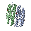


 Mass: 55.845 Da / Num. of mol.: 5 / Source method: obtained synthetically / Formula: Fe
 Mass: 65.409 Da / Num. of mol.: 6 / Source method: obtained synthetically / Formula: Zn
 Mass: 22.990 Da / Num. of mol.: 1 / Source method: obtained synthetically / Formula: Na
 Mass: 59.044 Da / Num. of mol.: 4 / Source method: obtained synthetically / Formula: C2H3O2
 Mass: 62.068 Da / Num. of mol.: 2 / Source method: obtained synthetically / Formula: C2H6O2
 Sample preparation
 / Beamline: 19-BM / Wavelength: 0.97883 Å
 Processing