 Movie
Movie Controller

[English] 日本語
 Yorodumi
- PDB-2gyk: Crystal structure of the complex of the Colicin E9 DNase domain w... -
+ Open data

- Basic information
| Entry | Database: PDB / ID: 2gyk | ||||||
|---|---|---|---|---|---|---|---|
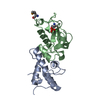
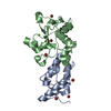
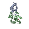
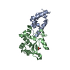
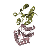
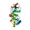
| Title | Crystal structure of the complex of the Colicin E9 DNase domain with a mutant immunity protein, IMME9 (D51A) | ||||||
 Components Components |
| ||||||
 Keywords Keywords | antibiotic/antibiotic inhibitor / PROTEIN-PROTEIN COMPLEX / 4-HELIX BUNDLE / DNASE DOMAIN / HNH-MOTIF / antibiotic-antibiotic inhibitor COMPLEX | ||||||
| Function / homology |  Function and homology information Function and homology informationextrachromosomal circular DNA / bacteriocin immunity / toxic substance binding / endonuclease activity / killing of cells of another organism / Hydrolases; Acting on ester bonds / defense response to bacterium / protein domain specific binding / hydrolase activity / protein-containing complex / metal ion binding Similarity search - Function | ||||||
| Biological species |  | ||||||
| Method |  X-RAY DIFFRACTION / SYNCHROTRON / MOLECULAR REPLACEMENT / Resolution: 1.6 Å X-RAY DIFFRACTION / SYNCHROTRON / MOLECULAR REPLACEMENT / Resolution: 1.6 Å | ||||||
 Authors Authors | Santi, P.S. / Kolade, O.O. / Kuhlmann, U.C. / Hemmings, A.M. | ||||||
 Citation Citation | Journal: To be Published Title: Crystal structures of the complexes of the Colicin E9 DNase domain with mutant immunity proteins Authors: Kuhlmann, U.C. / Santi, P.S. / Kolade, O.O. / Moore, G.R. / James, R. / Kleanthous, C. / Hemmings, A.M. #1: Journal: Nat.Struct.Biol. / Year: 1999 Title: Structural and mechanistic basis of immunity towards endonuclease colicins Authors: Kleanthous, C. / Kuhlmann, U.C. / Pommer, A.J. / Ferguson, N. / Radford, S.E. / Moore, G.R. / James, R. / Hemmings, A.M. | ||||||
| History |
|
- Structure visualization
| Structure viewer | Molecule: MolmilJmol/JSmol |
|---|
- Downloads & links
-Download
| PDBx/mmCIF format | 2gyk.cif.gz | 114.8 KB | Display | PDBx/mmCIF format |
|---|---|---|---|---|
| PDB format | pdb2gyk.ent.gz | 87.2 KB | Display | PDB format |
| PDBx/mmJSON format | 2gyk.json.gz | Tree view | PDBx/mmJSON format | |
| Others |  Other downloads Other downloads |
-Validation report
| Arichive directory | https://data.pdbj.org/pub/pdb/validation_reports/gy/2gykftp://data.pdbj.org/pub/pdb/validation_reports/gy/2gyk | HTTPS FTP |
|---|
-Related structure data
| Related structure data | 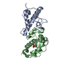 1emvS S: Starting model for refinement |
|---|---|
| Similar structure data |


-Links
 PDBj
PDBj- Assembly
| Deposited unit | 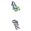
| ||||||||
|---|---|---|---|---|---|---|---|---|---|
| 1 | 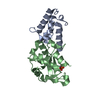
| ||||||||
| 2 | 
| ||||||||
| Unit cell |
| ||||||||
| Details | THE CRYSTALLOGRAPHIC ASYMMETRIC UNIT CONTAINS A DIMER OF THE BIOLOGICAL UNIT |
-Components
| #1: Protein | Mass: 9548.490 Da / Num. of mol.: 2 / Mutation: D51A Source method: isolated from a genetically manipulated source Source: (gene. exp.) #2: Protein | Mass: 15120.021 Da / Num. of mol.: 2 / Fragment: C-TERMINAL DOMAIN, DNASE DOMAIN Source method: isolated from a genetically manipulated source Source: (gene. exp.) #3: Chemical |   Mass: 65.409 Da / Num. of mol.: 2 / Source method: obtained synthetically / Formula: Zn Mass: 65.409 Da / Num. of mol.: 2 / Source method: obtained synthetically / Formula: Zn#4: Chemical |   Mass: 94.971 Da / Num. of mol.: 2 / Source method: obtained synthetically / Formula: PO4 Mass: 94.971 Da / Num. of mol.: 2 / Source method: obtained synthetically / Formula: PO4#5: Water | ChemComp-HOH / |  Mass: 18.015 Da / Num. of mol.: 686 / Source method: isolated from a natural source / Formula: H2O Mass: 18.015 Da / Num. of mol.: 686 / Source method: isolated from a natural source / Formula: H2O |
|---|
-Experimental details
-Experiment
| Experiment | Method: X-RAY DIFFRACTION / Number of used crystals: 1 |
|---|
- Sample preparation
| Crystal | Density Matthews: 2.1 Å3/Da / Density % sol: 41.3 % |
|---|---|
| Crystal grow | Temperature: 277 K / Method: vapor diffusion, sitting drop / pH: 5.3 Details: 24% (W/V) PEG 4K, 100mM SODIUM ACETATE BUFFER pH 5.3, VAPOR DIFFUSION, SITTING DROP, temperature 277K Temp details: 277 |
-Data collection
| Diffraction | Mean temperature: 100 K |
|---|---|
| Diffraction source | Source: SYNCHROTRON / Site: SRS  / Beamline: PX9.6 / Wavelength: 0.87 Å / Beamline: PX9.6 / Wavelength: 0.87 Å |
| Detector | Type: ADSC QUANTUM 4 / Detector: CCD / Date: Jun 3, 2000 |
| Radiation | Monochromator: SILICON(111) / Protocol: SINGLE WAVELENGTH / Monochromatic (M) / Laue (L): M / Scattering type: x-ray |
| Radiation wavelength | Wavelength: 0.87 Å / Relative weight: 1 |
| Reflection | Resolution: 1.6→20 Å / Num. all: 51260 / Num. obs: 51260 / % possible obs: 96.3 % / Observed criterion σ(F): 1 / Observed criterion σ(I): 1 / Rmerge(I) obs: 0.027 / Χ2: 1.422 / Net I/σ(I): 34.4 |
| Reflection shell | Resolution: 1.6→1.66 Å / Rmerge(I) obs: 0.045 / Num. unique all: 4468 / Χ2: 0.593 / % possible all: 84.5 |
- Processing
| Software |
| |||||||||||||||||||||||||||||||||||||||||||||||||||||||||||||||||||||||||||||||||||||||||||||||
|---|---|---|---|---|---|---|---|---|---|---|---|---|---|---|---|---|---|---|---|---|---|---|---|---|---|---|---|---|---|---|---|---|---|---|---|---|---|---|---|---|---|---|---|---|---|---|---|---|---|---|---|---|---|---|---|---|---|---|---|---|---|---|---|---|---|---|---|---|---|---|---|---|---|---|---|---|---|---|---|---|---|---|---|---|---|---|---|---|---|---|---|---|---|---|---|---|
| Refinement | Method to determine structure: MOLECULAR REPLACEMENT Starting model: PDB ENTRY 1EMV Resolution: 1.6→19.86 Å / Cor.coef. Fo:Fc: 0.953 / Cor.coef. Fo:Fc free: 0.929 / SU B: 3.39 / SU ML: 0.065 / Cross valid method: THROUGHOUT / σ(F): 3 / ESU R: 0.109 / ESU R Free: 0.111 / Stereochemistry target values: Engh & Huber / Details: HYDROGENS HAVE BEEN ADDED IN THE RIDING POSITIONS
| |||||||||||||||||||||||||||||||||||||||||||||||||||||||||||||||||||||||||||||||||||||||||||||||
| Solvent computation | Ion probe radii: 0.8 Å / Shrinkage radii: 0.8 Å / VDW probe radii: 1.2 Å / Solvent model: MASK | |||||||||||||||||||||||||||||||||||||||||||||||||||||||||||||||||||||||||||||||||||||||||||||||
| Displacement parameters | Biso mean: 15.426 Å2
| |||||||||||||||||||||||||||||||||||||||||||||||||||||||||||||||||||||||||||||||||||||||||||||||
| Refinement step | Cycle: LAST / Resolution: 1.6→19.86 Å
| |||||||||||||||||||||||||||||||||||||||||||||||||||||||||||||||||||||||||||||||||||||||||||||||
| Refine LS restraints |
| |||||||||||||||||||||||||||||||||||||||||||||||||||||||||||||||||||||||||||||||||||||||||||||||
| LS refinement shell | Resolution: 1.6→1.644 Å / Total num. of bins used: 20
|
