 Movie
Movie Controller

[English] 日本語
 Yorodumi
- PDB-2gwq: Crystal structure of D(G4T4G4) with four quadruplexes in the asym... -
+ Open data

- Basic information
| Entry | Database: PDB / ID: 2gwq | ||||||||||||||||||
|---|---|---|---|---|---|---|---|---|---|---|---|---|---|---|---|---|---|---|---|
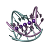
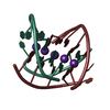
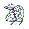
| Title | Crystal structure of D(G4T4G4) with four quadruplexes in the asymmetric unit. | ||||||||||||||||||
 Components Components | 5'-D(* Keywords KeywordsDNA / OXYTRICHA / G-QUARTET / G-QUADRUPLEX / G-TETRAD / G-TETRAPLEX / FOUR QUADRUPLEXES / ASYMMETRIC UNIT. | Function / homology | : / DNA / DNA (> 10) |  Function and homology information Function and homology informationMethod |  X-RAY DIFFRACTION / SYNCHROTRON / MOLECULAR REPLACEMENT / Resolution: 2 Å X-RAY DIFFRACTION / SYNCHROTRON / MOLECULAR REPLACEMENT / Resolution: 2 Å  Authors AuthorsLee, M.P.H. / Parkinson, G.N. / Neidle, S. |  CitationJournal: To be Published CitationJournal: To be PublishedTitle: Crystal structure of D(G4T4G4) with four and six quadruplexes in the asymmetric unit. Authors: Lee, M.P.H. / Haider, S. / Parkinson, G.N. / Neidle, S. History |
|
- Structure visualization
| Structure viewer | Molecule: MolmilJmol/JSmol |
|---|
- Downloads & links
-Download
| PDBx/mmCIF format | 2gwq.cif.gz | 73.4 KB | Display | PDBx/mmCIF format |
|---|---|---|---|---|
| PDB format | pdb2gwq.ent.gz | 53.2 KB | Display | PDB format |
| PDBx/mmJSON format | 2gwq.json.gz | Tree view | PDBx/mmJSON format | |
| Others |  Other downloads Other downloads |
-Validation report
| Arichive directory | https://data.pdbj.org/pub/pdb/validation_reports/gw/2gwqftp://data.pdbj.org/pub/pdb/validation_reports/gw/2gwq | HTTPS FTP |
|---|
-Related structure data
| Related structure data |  2gweC 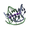 1jrnS S: Starting model for refinement C: citing same article ( |
|---|---|
| Similar structure data |


-Links
 PDBj
PDBj


- Assembly
| Deposited unit | 
| ||||||||
|---|---|---|---|---|---|---|---|---|---|
| 1 | 
| ||||||||
| 2 | 
| ||||||||
| 3 | 
| ||||||||
| 4 | 
| ||||||||
| Unit cell |
|
-Components
| #1: DNA chain | Mass: 3805.460 Da / Num. of mol.: 8 / Source method: obtained synthetically / Details: OXYTRICHA NOVA TELOMERIC SEQUENCE #2: Chemical | ChemComp-K /   Mass: 39.098 Da / Num. of mol.: 20 / Source method: obtained synthetically / Formula: K Mass: 39.098 Da / Num. of mol.: 20 / Source method: obtained synthetically / Formula: K#3: Water | ChemComp-HOH / |  Mass: 18.015 Da / Num. of mol.: 169 / Source method: isolated from a natural source / Formula: H2O Mass: 18.015 Da / Num. of mol.: 169 / Source method: isolated from a natural source / Formula: H2O |
|---|
-Experimental details
-Experiment
| Experiment | Method: X-RAY DIFFRACTION / Number of used crystals: 1 |
|---|
- Sample preparation
| Crystal | Density Matthews: 2.21 Å3/Da / Density % sol: 44.3 % | ||||||||||||||||||||||||||||||||||||||||||||
|---|---|---|---|---|---|---|---|---|---|---|---|---|---|---|---|---|---|---|---|---|---|---|---|---|---|---|---|---|---|---|---|---|---|---|---|---|---|---|---|---|---|---|---|---|---|
| Crystal grow | Temperature: 285 K / pH: 6.5 Details: MAGNESIUM CHLORIDE, DNA, POTASSIUM CHLORIDE, MPD, PEPTIDE CONJUGATE BIS RKKV ACRIDONE, SPERMINE TETRAHYDROCHLORIDE, POTASSIUM CACODYLATE BUFFER, VAPOR DIFFUSION, HANGING DROP, temperature 285K, pH 6.50 | ||||||||||||||||||||||||||||||||||||||||||||
| Components of the solutions |
|
-Data collection
| Diffraction | Mean temperature: 100 K |
|---|---|
| Diffraction source | Source: SYNCHROTRON / Site: ESRF  / Beamline: ID29 / Wavelength: 0.862451 / Beamline: ID29 / Wavelength: 0.862451 |
| Detector | Type: ADSC QUANTUM 210 / Detector: CCD / Date: Apr 28, 2003 / Details: A CYLINDRICAL GRAZING INCIDENCE MIRROR |
| Radiation | Monochromator: LIQUID NITROGEN COOLED CHANNEL-CUT SILICON MONOCHROMATOR Protocol: SINGLE WAVELENGTH / Monochromatic (M) / Laue (L): M / Scattering type: x-ray |
| Radiation wavelength | Wavelength: 0.862451 Å / Relative weight: 1 |
| Reflection | Resolution: 2→17 Å / Num. obs: 18992 / % possible obs: 96.6 % / Observed criterion σ(I): -2 / Redundancy: 3.35 % / Rmerge(I) obs: 0.091 / Net I/σ(I): 26.27 |
| Reflection shell | Resolution: 2→2.07 Å / Rmerge(I) obs: 0.197 / Mean I/σ(I) obs: 8.74 / % possible all: 96.8 |
- Processing
| Software |
| |||||||||||||||||||||||||||||||||
|---|---|---|---|---|---|---|---|---|---|---|---|---|---|---|---|---|---|---|---|---|---|---|---|---|---|---|---|---|---|---|---|---|---|---|
| Refinement | Method to determine structure: MOLECULAR REPLACEMENT Starting model: DNA PART OF NDB ENTRY CODE UD0014 OR PDB ENTRY CODE 1JRN. Resolution: 2→8 Å / Num. parameters: 8867 / Num. restraintsaints: 10018 / Cross valid method: THROUGHOUT / σ(F): 4 / Stereochemistry target values: ENGH & HUBER Details: ANISOTROPIC SCALING APPLIED BY THE METHOD OF PARKIN, MOEZZI & HOPE, J.APPL.CRYST.28(1995)53-56
| |||||||||||||||||||||||||||||||||
| Solvent computation | Solvent model: MOEWS & KRETSINGER, J.MOL.BIOL.91(1973)201-228 | |||||||||||||||||||||||||||||||||
| Displacement parameters | Biso mean: 17.84 Å2 | |||||||||||||||||||||||||||||||||
| Refine analyze | Num. disordered residues: 0 / Occupancy sum hydrogen: 0 / Occupancy sum non hydrogen: 2213 | |||||||||||||||||||||||||||||||||
| Refinement step | Cycle: LAST / Resolution: 2→8 Å
| |||||||||||||||||||||||||||||||||
| Refine LS restraints |
|
