 Movie
Movie Controller

[English] 日本語
 Yorodumi
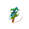
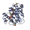
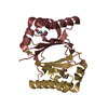
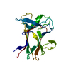
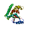
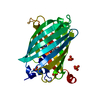
- PDB-2gu9: Crystal structure of XC5357 from Xanthomonas campestris: A putati... -
+ Open data

- Basic information
| Entry | Database: PDB / ID: 2gu9 | ||||||
|---|---|---|---|---|---|---|---|
| Title | Crystal structure of XC5357 from Xanthomonas campestris: A putative tetracenomycin polyketide synthesis protein adopting a novel cupin subfamily structure | ||||||
 Components Components | tetracenomycin polyketide synthesis protein | ||||||
 Keywords Keywords | IMMUNE SYSTEM / Xanthomonas campestris / cupin / tetracenomycin polyketide | ||||||
| Function / homology |  Function and homology information Function and homology information | ||||||
| Biological species |  Xanthomonas campestris (bacteria) Xanthomonas campestris (bacteria) | ||||||
| Method |  X-RAY DIFFRACTION / SYNCHROTRON / MAD / Resolution: 1.4 Å X-RAY DIFFRACTION / SYNCHROTRON / MAD / Resolution: 1.4 Å | ||||||
 Authors Authors | Chin, K.-H. / Chou, C.C. / Wang, A.H.-J. / Chou, S.-H. | ||||||
 Citation Citation | Journal: Proteins / Year: 2006 Title: Crystal structure of XC5357 from Xanthomonas campestris: A putative tetracenomycin polyketide synthesis protein adopting a novel cupin subfamily structure Authors: Chin, K.-H. / Chou, C.C. / Wang, A.H.-J. / Chou, S.-H. | ||||||
| History |
|
- Structure visualization
| Structure viewer | Molecule: MolmilJmol/JSmol |
|---|
- Downloads & links
-Download
| PDBx/mmCIF format | 2gu9.cif.gz | 50.2 KB | Display | PDBx/mmCIF format |
|---|---|---|---|---|
| PDB format | pdb2gu9.ent.gz | 37.4 KB | Display | PDB format |
| PDBx/mmJSON format | 2gu9.json.gz | Tree view | PDBx/mmJSON format | |
| Others |  Other downloads Other downloads |
-Validation report
| Summary document | 2gu9_validation.pdf.gz | 370.7 KB | Display | wwPDB validaton report |
|---|---|---|---|---|
| Full document | 2gu9_full_validation.pdf.gz | 372.5 KB | Display | |
| Data in XML | 2gu9_validation.xml.gz | 5.6 KB | Display | |
| Data in CIF | 2gu9_validation.cif.gz | 8.2 KB | Display | |
| Arichive directory | https://data.pdbj.org/pub/pdb/validation_reports/gu/2gu9ftp://data.pdbj.org/pub/pdb/validation_reports/gu/2gu9 | HTTPS FTP |
-Related structure data
| Similar structure data |
|---|


-Links
 PDBj
PDBj- Assembly
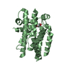
| Deposited unit | 
| ||||||||
|---|---|---|---|---|---|---|---|---|---|
| 1 |
| ||||||||
| Unit cell |
|
-Components
| #1: Protein | Mass: 12178.454 Da / Num. of mol.: 2 Source method: isolated from a genetically manipulated source Source: (gene. exp.) Xanthomonas campestris (bacteria) / Plasmid: pET30 / Species (production host): Escherichia coli / Production host: |
|---|
-Experimental details
-Experiment
| Experiment | Method: X-RAY DIFFRACTION / Number of used crystals: 12 |
|---|
- Sample preparation
| Crystal | Density Matthews: 2.96 Å3/Da / Density % sol: 58.5 % |
|---|---|
| Crystal grow | Temperature: 293 K / Method: vapor diffusion, hanging drop / pH: 5.6 Details: 0.1M NaOAC, 1.6M LiSO4, pH 5.6, VAPOR DIFFUSION, HANGING DROP, temperature 293K |
-Data collection
| Diffraction |
| ||||||||||||||||||
|---|---|---|---|---|---|---|---|---|---|---|---|---|---|---|---|---|---|---|---|
| Diffraction source |
| ||||||||||||||||||
| Detector |
| ||||||||||||||||||
| Radiation |
| ||||||||||||||||||
| Radiation wavelength |
| ||||||||||||||||||
| Reflection | Resolution: 1.4→30 Å / Num. all: 33553 / Num. obs: 32312 / % possible obs: 96.3 % / Observed criterion σ(F): 2 / Observed criterion σ(I): 2 | ||||||||||||||||||
| Reflection shell | Resolution: 1.4→1.46 Å / % possible all: 96.3 |

- Processing
| Software |
| ||||||||||||||||||||
|---|---|---|---|---|---|---|---|---|---|---|---|---|---|---|---|---|---|---|---|---|---|
| Refinement | Method to determine structure: MAD / Resolution: 1.4→30 Å / Cross valid method: THROUGHOUT / σ(F): 2 / Stereochemistry target values: Engh & Huber
| ||||||||||||||||||||
| Refinement step | Cycle: LAST / Resolution: 1.4→30 Å
| ||||||||||||||||||||
| Refine LS restraints | Type: c_angle_deg / Dev ideal: 0.02 |
