- PDB-2gu1: Crystal structure of a zinc containing peptidase from vibrio cholerae -
+
Open data
ID or keywords:
Loading...
-
Basic information
Entry
Database: PDB / ID: 2gu1
Title
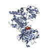
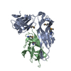
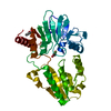
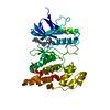
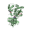
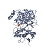
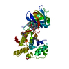
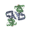
Crystal structure of a zinc containing peptidase from vibrio cholerae
Components
Zinc peptidase
Keywords
HYDROLASE / Zinc Peptidase / alpha/beta / beta barrel / Structural Genomics / PSI / Protein Structure Initiative / New York SGX Research Center for Structural Genomics / NYSGXRC
Function / homology
Function and homology information
peptidoglycan binding / Hydrolases; Acting on peptide bonds (peptidases); Metalloendopeptidases / cell wall organization / metalloendopeptidase activity / periplasmic space / proteolysis / metal ion binding Similarity search - Function
Opacity-associated protein A / Opacity-associated protein A LysM-like domain / Nuclear Transport Factor 2; Chain: A, - #350 / Csd3-like, second N-terminal domain / Csd3 second domain / Glucose Permease (Domain IIA) / Glucose Permease (Domain IIA) / : / Peptidase M23 / Peptidase family M23 ...Opacity-associated protein A / Opacity-associated protein A LysM-like domain / Nuclear Transport Factor 2; Chain: A, - #350 / Csd3-like, second N-terminal domain / Csd3 second domain / Glucose Permease (Domain IIA) / Glucose Permease (Domain IIA) / : / Peptidase M23 / Peptidase family M23 / Duplicated hybrid motif / Nuclear Transport Factor 2; Chain: A, / Distorted Sandwich / Roll / Mainly Beta / Alpha Beta Similarity search - Domain/homology
Method to determine structure: MAD / Resolution: 1.9→32.24 Å / Rfactor Rfree error: 0.006 / Data cutoff high absF: 142984.74 / Data cutoff low absF: 0 / Isotropic thermal model: RESTRAINED / Cross valid method: THROUGHOUT / σ(F): 0 / Stereochemistry target values: Engh & Huber Details: Missing residues listed in remark 465 were not modeled due to lack of electron density. Atoms missing in some residues listed in remark 470 are due to lack of density for side chain atoms ...Details: Missing residues listed in remark 465 were not modeled due to lack of electron density. Atoms missing in some residues listed in remark 470 are due to lack of density for side chain atoms and the relevant residues were modeled as ALA.
In the structure databanks used in Yorodumi, some data are registered as the other names, "COVID-19 virus" and "2019-nCoV". Here are the details of the virus and the list of structure data.
Jan 31, 2019. EMDB accession codes are about to change! (news from PDBe EMDB page)
EMDB accession codes are about to change! (news from PDBe EMDB page)
The allocation of 4 digits for EMDB accession codes will soon come to an end. Whilst these codes will remain in use, new EMDB accession codes will include an additional digit and will expand incrementally as the available range of codes is exhausted. The current 4-digit format prefixed with “EMD-” (i.e. EMD-XXXX) will advance to a 5-digit format (i.e. EMD-XXXXX), and so on. It is currently estimated that the 4-digit codes will be depleted around Spring 2019, at which point the 5-digit format will come into force.
The EM Navigator/Yorodumi systems omit the EMD- prefix.
Related info.:Q: What is EMD? / ID/Accession-code notation in Yorodumi/EM Navigator
Yorodumi is a browser for structure data from EMDB, PDB, SASBDB, etc.
This page is also the successor to EM Navigator detail page, and also detail information page/front-end page for Omokage search.
The word "yorodu" (or yorozu) is an old Japanese word meaning "ten thousand". "mi" (miru) is to see.
Related info.:EMDB / PDB / SASBDB / Comparison of 3 databanks / Yorodumi Search / Aug 31, 2016. New EM Navigator & Yorodumi / Yorodumi Papers / Jmol/JSmol / Function and homology information / Changes in new EM Navigator and Yorodumi
 Movie
Movie Controller

 Yorodumi
 Open data
 Basic information
 Components
 Keywords
 Function and homology information
 Vibrio cholerae (bacteria)
 X-RAY DIFFRACTION /
  Authors
 Citation
 Structure visualization
 Downloads & links
 Other downloads

 PDBj
PDBj Assembly
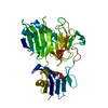

 Mass: 65.409 Da / Num. of mol.: 1 / Source method: obtained synthetically / Formula: Zn
 Mass: 22.990 Da / Num. of mol.: 1 / Source method: obtained synthetically / Formula: Na
 Mass: 18.015 Da / Num. of mol.: 133 / Source method: isolated from a natural source / Formula: H2O
 Sample preparation
 Processing