 Movie
Movie Controller

[English] 日本語
 Yorodumi
- PDB-2goj: The crystal structure of the enzyme Fe-superoxide dismutase from ... -
+ Open data

- Basic information
| Entry | Database: PDB / ID: 2goj | ||||||
|---|---|---|---|---|---|---|---|
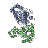
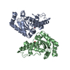
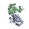
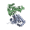
| Title | The crystal structure of the enzyme Fe-superoxide dismutase from Plasmodium falciparum | ||||||
 Components Components | Fe-superoxide dismutase | ||||||
 Keywords Keywords | OXIDOREDUCTASE / BETA+ALPHA STRUCTURE | ||||||
| Function / homology |  Function and homology information Function and homology informationsuperoxide dismutase / superoxide dismutase activity / metal ion binding / cytoplasm Similarity search - Function | ||||||
| Biological species |  | ||||||
| Method |  X-RAY DIFFRACTION / MOLECULAR REPLACEMENT / Resolution: 2 Å X-RAY DIFFRACTION / MOLECULAR REPLACEMENT / Resolution: 2 Å | ||||||
 Authors Authors | Navarro, M.V.A.S. / Garratt, R.C. | ||||||
 Citation Citation | Journal: Proteins / Year: 2009 Title: Systematic structural studies of iron superoxide dismutases from human parasites and a statistical coupling analysis of metal binding specificity Authors: Bachega, J.F. / Navarro, M.V. / Bleicher, L. / Bortoleto-Bugs, R.K. / Dive, D. / Hoffmann, P. / Viscogliosi, E. / Garratt, R.C. | ||||||
| History |
|
- Structure visualization
| Structure viewer | Molecule: MolmilJmol/JSmol |
|---|
- Downloads & links
-Download
| PDBx/mmCIF format | 2goj.cif.gz | 96.4 KB | Display | PDBx/mmCIF format |
|---|---|---|---|---|
| PDB format | pdb2goj.ent.gz | 74.1 KB | Display | PDB format |
| PDBx/mmJSON format | 2goj.json.gz | Tree view | PDBx/mmJSON format | |
| Others |  Other downloads Other downloads |
-Validation report
| Arichive directory | https://data.pdbj.org/pub/pdb/validation_reports/go/2gojftp://data.pdbj.org/pub/pdb/validation_reports/go/2goj | HTTPS FTP |
|---|
-Related structure data
| Related structure data |  2gpcC  3esfC  1isaS S: Starting model for refinement C: citing same article ( |
|---|---|
| Similar structure data |


-Links
 PDBj
PDBj


- Assembly
| Deposited unit | 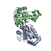
| ||||||||
|---|---|---|---|---|---|---|---|---|---|
| 1 |
| ||||||||
| Unit cell |
| ||||||||
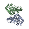
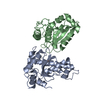
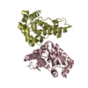
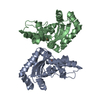
| Details | The assymetric unit contains a dimer, which represents the molecular assembly |
-Components
| #1: Protein | Mass: 22630.441 Da / Num. of mol.: 2 Source method: isolated from a genetically manipulated source Source: (gene. exp.) Strain: HB3 / Gene: SOD / Species (production host): Escherichia coli / Production host:  References: GenBank: 861077, UniProt: Q27740*PLUS, superoxide dismutase #2: Chemical |   Mass: 55.845 Da / Num. of mol.: 2 / Source method: obtained synthetically / Formula: Fe Mass: 55.845 Da / Num. of mol.: 2 / Source method: obtained synthetically / Formula: Fe#3: Water | ChemComp-HOH / |  Mass: 18.015 Da / Num. of mol.: 305 / Source method: isolated from a natural source / Formula: H2O Mass: 18.015 Da / Num. of mol.: 305 / Source method: isolated from a natural source / Formula: H2O |
|---|
-Experimental details
-Experiment
| Experiment | Method: X-RAY DIFFRACTION / Number of used crystals: 1 |
|---|
- Sample preparation
| Crystal | Density Matthews: 2.12 Å3/Da / Density % sol: 41.98 % |
|---|---|
| Crystal grow | Temperature: 291 K / Method: vapor diffusion, hanging drop / pH: 8.5 Details: 25.5% PEG 4000 (w/v), 0.17 M Sodium Acetate, 0.085 M Tris-HCl, pH 8.5, VAPOR DIFFUSION, HANGING DROP, temperature 291K |
-Data collection
| Diffraction | Mean temperature: 100 K |
|---|---|
| Diffraction source | Source: ROTATING ANODE / Type: RIGAKU / Wavelength: 1.5418 Å |
| Detector | Type: RIGAKU RAXIS IIC / Detector: IMAGE PLATE / Date: Feb 10, 2002 |
| Radiation | Monochromator: GRAPHITE / Protocol: SINGLE WAVELENGTH / Monochromatic (M) / Laue (L): M / Scattering type: x-ray |
| Radiation wavelength | Wavelength: 1.5418 Å / Relative weight: 1 |
| Reflection | Resolution: 2→31.62 Å / Num. obs: 25131 / % possible obs: 95.3 % / Observed criterion σ(F): 2 / Observed criterion σ(I): 2 / Redundancy: 6.4 % / Rsym value: 0.098 / Net I/σ(I): 7.5 |
| Reflection shell | Resolution: 2→2.11 Å / Redundancy: 2.8 % / Mean I/σ(I) obs: 2 / Rsym value: 0.37 / % possible all: 73.3 |
- Processing
| Software | Name: REFMAC / Version: 5.1.24 / Classification: refinement | |||||||||||||||||||||||||||||||||||||||||||||||||||||||||||||||||||||||||||||||||||||||||||||||||||||||||
|---|---|---|---|---|---|---|---|---|---|---|---|---|---|---|---|---|---|---|---|---|---|---|---|---|---|---|---|---|---|---|---|---|---|---|---|---|---|---|---|---|---|---|---|---|---|---|---|---|---|---|---|---|---|---|---|---|---|---|---|---|---|---|---|---|---|---|---|---|---|---|---|---|---|---|---|---|---|---|---|---|---|---|---|---|---|---|---|---|---|---|---|---|---|---|---|---|---|---|---|---|---|---|---|---|---|---|
| Refinement | Method to determine structure: MOLECULAR REPLACEMENT Starting model: PDB ENTRY 1ISA Resolution: 2→31.62 Å / Cor.coef. Fo:Fc: 0.955 / Cor.coef. Fo:Fc free: 0.943 / SU B: 3.711 / SU ML: 0.101 / Cross valid method: THROUGHOUT / σ(F): 2 / ESU R: 0.191 / ESU R Free: 0.153 / Stereochemistry target values: MAXIMUM LIKELIHOOD / Details: HYDROGENS HAVE BEEN ADDED IN THE RIDING POSITIONS
| |||||||||||||||||||||||||||||||||||||||||||||||||||||||||||||||||||||||||||||||||||||||||||||||||||||||||
| Solvent computation | Ion probe radii: 0.8 Å / Shrinkage radii: 0.8 Å / VDW probe radii: 1.4 Å / Solvent model: BABINET MODEL WITH MASK | |||||||||||||||||||||||||||||||||||||||||||||||||||||||||||||||||||||||||||||||||||||||||||||||||||||||||
| Displacement parameters | Biso mean: 14.498 Å2
| |||||||||||||||||||||||||||||||||||||||||||||||||||||||||||||||||||||||||||||||||||||||||||||||||||||||||
| Refinement step | Cycle: LAST / Resolution: 2→31.62 Å
| |||||||||||||||||||||||||||||||||||||||||||||||||||||||||||||||||||||||||||||||||||||||||||||||||||||||||
| Refine LS restraints |
| |||||||||||||||||||||||||||||||||||||||||||||||||||||||||||||||||||||||||||||||||||||||||||||||||||||||||
| LS refinement shell | Resolution: 2→2.108 Å / Total num. of bins used: 10 /
|
