 Movie
Movie Controller

[English] 日本語
 Yorodumi
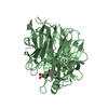
- PDB-2gh6: Crystal structure of a HDAC-like protein with 9,9,9-trifluoro-8-o... -
+ Open data

- Basic information
| Entry | Database: PDB / ID: 2gh6 | ||||||
|---|---|---|---|---|---|---|---|
| Title | Crystal structure of a HDAC-like protein with 9,9,9-trifluoro-8-oxo-N-phenylnonan amide bound | ||||||
 Components Components | Histone deacetylase-like amidohydrolase | ||||||
 Keywords Keywords | HYDROLASE / Histone deacetylase / zinc-ion / trifluoromethyl ketone | ||||||
| Function / homology |  Function and homology information Function and homology informationhistone deacetylase activity / Hydrolases; Acting on carbon-nitrogen bonds, other than peptide bonds; In linear amides / epigenetic regulation of gene expression / hydrolase activity / metal ion binding Similarity search - Function | ||||||
| Biological species |  Alcaligenaceae bacterium FB188 (bacteria) Alcaligenaceae bacterium FB188 (bacteria) | ||||||
| Method |  X-RAY DIFFRACTION / MOLECULAR REPLACEMENT / Resolution: 2.203 Å X-RAY DIFFRACTION / MOLECULAR REPLACEMENT / Resolution: 2.203 Å | ||||||
 Authors Authors | Nielsen, T.K. / Hildmann, C. / Riester, D. / Wegener, D. / Schwienhorst, A. / Ficner, R. | ||||||
 Citation Citation | Journal: Acta Crystallogr.,Sect.F / Year: 2007 Title: Complex structure of a bacterial class 2 histone deacetylase homologue with a trifluoromethylketone inhibitor. Authors: Nielsen, T.K. / Hildmann, C. / Riester, D. / Wegener, D. / Schwienhorst, A. / Ficner, R. | ||||||
| History |
|
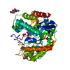
- Structure visualization
| Structure viewer | Molecule: MolmilJmol/JSmol |
|---|
- Downloads & links
-Download
| PDBx/mmCIF format | 2gh6.cif.gz | 299.9 KB | Display | PDBx/mmCIF format |
|---|---|---|---|---|
| PDB format | pdb2gh6.ent.gz | 240.5 KB | Display | PDB format |
| PDBx/mmJSON format | 2gh6.json.gz | Tree view | PDBx/mmJSON format | |
| Others |  Other downloads Other downloads |
-Validation report
| Arichive directory | https://data.pdbj.org/pub/pdb/validation_reports/gh/2gh6ftp://data.pdbj.org/pub/pdb/validation_reports/gh/2gh6 | HTTPS FTP |
|---|
-Related structure data
| Related structure data |  1zz1S S: Starting model for refinement |
|---|---|
| Similar structure data |


-Links
 PDBj
PDBj- Assembly
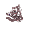
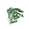
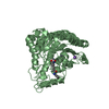
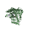
| Deposited unit | 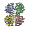
| ||||||||||||||||||||||||||||||
|---|---|---|---|---|---|---|---|---|---|---|---|---|---|---|---|---|---|---|---|---|---|---|---|---|---|---|---|---|---|---|---|
| 1 | 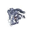
| ||||||||||||||||||||||||||||||
| 2 | 
| ||||||||||||||||||||||||||||||
| 3 | 
| ||||||||||||||||||||||||||||||
| 4 | 
| ||||||||||||||||||||||||||||||
| 5 |
| ||||||||||||||||||||||||||||||
| Unit cell |
| ||||||||||||||||||||||||||||||
| Noncrystallographic symmetry (NCS) | NCS domain:
NCS domain segments: Component-ID: 1 / Ens-ID: 1 / Beg auth comp-ID: ALA / Beg label comp-ID: ALA / End auth comp-ID: ILE / End label comp-ID: ILE / Refine code: 5 / Auth seq-ID: 2 - 368 / Label seq-ID: 2 - 368
| ||||||||||||||||||||||||||||||
| Details | Protein is a monomer |
-Components
| #1: Protein | Mass: 39424.602 Da / Num. of mol.: 4 Source method: isolated from a genetically manipulated source Source: (gene. exp.) Alcaligenaceae bacterium FB188 (bacteria)Strain: DSM 11172 Description: source organism synonym Bordetella sp. (strain FB188) Gene: hdaH, hdaH1 / Plasmid: pQE / Production host: References: UniProt: Q70I53, Hydrolases; Acting on carbon-nitrogen bonds, other than peptide bonds; In linear amides #2: Chemical | ChemComp-ZN /   Mass: 65.409 Da / Num. of mol.: 4 / Source method: obtained synthetically / Formula: Zn Mass: 65.409 Da / Num. of mol.: 4 / Source method: obtained synthetically / Formula: Zn#3: Chemical | ChemComp-K /   Mass: 39.098 Da / Num. of mol.: 8 / Source method: obtained synthetically / Formula: K Mass: 39.098 Da / Num. of mol.: 8 / Source method: obtained synthetically / Formula: K#4: Chemical | ChemComp-CF3 /   Mass: 301.304 Da / Num. of mol.: 4 / Source method: obtained synthetically / Formula: C15H18F3NO2 Mass: 301.304 Da / Num. of mol.: 4 / Source method: obtained synthetically / Formula: C15H18F3NO2#5: Water | ChemComp-HOH / |  Mass: 18.015 Da / Num. of mol.: 783 / Source method: isolated from a natural source / Formula: H2O Mass: 18.015 Da / Num. of mol.: 783 / Source method: isolated from a natural source / Formula: H2O |
|---|
-Experimental details
-Experiment
| Experiment | Method: X-RAY DIFFRACTION / Number of used crystals: 1 |
|---|
- Sample preparation
| Crystal | Density Matthews: 2.46 Å3/Da / Density % sol: 50.03 % |
|---|---|
| Crystal grow | Temperature: 293 K / Method: vapor diffusion, sitting drop / pH: 6.5 Details: NaCl, Na-cacodylate, pH 6.5, VAPOR DIFFUSION, SITTING DROP, temperature 293K |
-Data collection
| Diffraction source | Source: ROTATING ANODE / Type: RIGAKU / Wavelength: 1.5418 Å |
|---|---|
| Detector | Type: RIGAKU / Detector: IMAGE PLATE |
| Radiation | Protocol: SINGLE WAVELENGTH / Monochromatic (M) / Laue (L): M / Scattering type: x-ray |
| Radiation wavelength | Wavelength: 1.5418 Å / Relative weight: 1 |
| Reflection | Resolution: 2.203→39.84 Å / Num. all: 76561 / Num. obs: 76561 |
| Reflection shell | Resolution: 2.203→2.26 Å / % possible all: 92.9 |
- Processing
| Software |
| ||||||||||||||||||||||||||||||||||||||||||||||||||||||||||||||||||||||||||||||||||||||||||||||||||||||
|---|---|---|---|---|---|---|---|---|---|---|---|---|---|---|---|---|---|---|---|---|---|---|---|---|---|---|---|---|---|---|---|---|---|---|---|---|---|---|---|---|---|---|---|---|---|---|---|---|---|---|---|---|---|---|---|---|---|---|---|---|---|---|---|---|---|---|---|---|---|---|---|---|---|---|---|---|---|---|---|---|---|---|---|---|---|---|---|---|---|---|---|---|---|---|---|---|---|---|---|---|---|---|---|
| Refinement | Method to determine structure: MOLECULAR REPLACEMENT Starting model: PDB ENTRY 1ZZ1 Resolution: 2.203→39.84 Å / Cor.coef. Fo:Fc: 0.96 / Cor.coef. Fo:Fc free: 0.931 / SU B: 5.015 / SU ML: 0.129 / Cross valid method: THROUGHOUT / σ(F): 0 / ESU R: 0.244 / ESU R Free: 0.195 / Stereochemistry target values: MAXIMUM LIKELIHOOD
| ||||||||||||||||||||||||||||||||||||||||||||||||||||||||||||||||||||||||||||||||||||||||||||||||||||||
| Solvent computation | Ion probe radii: 0.8 Å / Shrinkage radii: 0.8 Å / VDW probe radii: 1.2 Å / Solvent model: MASK | ||||||||||||||||||||||||||||||||||||||||||||||||||||||||||||||||||||||||||||||||||||||||||||||||||||||
| Displacement parameters | Biso mean: 24.778 Å2
| ||||||||||||||||||||||||||||||||||||||||||||||||||||||||||||||||||||||||||||||||||||||||||||||||||||||
| Refinement step | Cycle: LAST / Resolution: 2.203→39.84 Å
| ||||||||||||||||||||||||||||||||||||||||||||||||||||||||||||||||||||||||||||||||||||||||||||||||||||||
| Refine LS restraints |
| ||||||||||||||||||||||||||||||||||||||||||||||||||||||||||||||||||||||||||||||||||||||||||||||||||||||
| Refine LS restraints NCS | Ens-ID: 1 / Refine-ID: X-RAY DIFFRACTION
| ||||||||||||||||||||||||||||||||||||||||||||||||||||||||||||||||||||||||||||||||||||||||||||||||||||||
| LS refinement shell | Resolution: 2.203→2.26 Å / Total num. of bins used: 20
|
