 Movie
Movie Controller

[English] 日本語
 Yorodumi
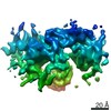
- PDB-2gag: Heteroteterameric sarcosine: structure of a diflavin metaloenzyme... -
+ Open data

- Basic information
| Entry | Database: PDB / ID: 2gag | ||||||
|---|---|---|---|---|---|---|---|
| Title | Heteroteterameric sarcosine: structure of a diflavin metaloenzyme at 1.85 a resolution | ||||||
 Components Components | (heterotetrameric sarcosine oxidase ...) x 4 | ||||||
 Keywords Keywords | OXIDOREDUCTASE / Sarcosine oxidase / Flavoenzyme / Electron transfer / Folate-methylating enzyme | ||||||
| Function / homology |  Function and homology information Function and homology informationsarcosine oxidase (5,10-methylenetetrahydrofolate-forming) / : / sarcosine oxidase activity / tetrahydrofolate metabolic process / transaminase activity / nucleotide binding / metal ion binding / cytoplasm Similarity search - Function | ||||||
| Biological species |  Stenotrophomonas maltophilia (bacteria) Stenotrophomonas maltophilia (bacteria) | ||||||
| Method |  X-RAY DIFFRACTION / SYNCHROTRON / MOLECULAR REPLACEMENT / Resolution: 1.85 Å X-RAY DIFFRACTION / SYNCHROTRON / MOLECULAR REPLACEMENT / Resolution: 1.85 Å | ||||||
 Authors Authors | Chen, Z.W. / Hassan-Abdulah, A. / Zhao, G. / Jorns, M.S. / Mathews, F.S. | ||||||
 Citation Citation | Journal: J.Mol.Biol. / Year: 2006 Title: Heterotetrameric sarcosine oxidase: structure of a diflavin metalloenzyme at 1.85 a resolution. Authors: Chen, Z.W. / Hassan-Abdulah, A. / Zhao, G. / Jorns, M.S. / Mathews, F.S. #1: Journal: Protein Expr.Purif. / Year: 2005 Title: Cloning, expression and crystallization of heterotetrameric sarcosine oxidase from Pseudomonas maltophilia | ||||||
| History |
|
- Structure visualization
| Structure viewer | Molecule: MolmilJmol/JSmol |
|---|
- Downloads & links
-Download
| PDBx/mmCIF format | 2gag.cif.gz | 365.7 KB | Display | PDBx/mmCIF format |
|---|---|---|---|---|
| PDB format | pdb2gag.ent.gz | 285 KB | Display | PDB format |
| PDBx/mmJSON format | 2gag.json.gz | Tree view | PDBx/mmJSON format | |
| Others |  Other downloads Other downloads |
-Validation report
| Arichive directory | https://data.pdbj.org/pub/pdb/validation_reports/ga/2gagftp://data.pdbj.org/pub/pdb/validation_reports/ga/2gag | HTTPS FTP |
|---|
-Related structure data
| Related structure data |  2gahSC S: Starting model for refinement C: citing same article ( |
|---|---|
| Similar structure data |

-Links
 PDBj
PDBj


- Assembly
| Deposited unit | 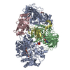
| ||||||||
|---|---|---|---|---|---|---|---|---|---|
| 1 |
| ||||||||
| Unit cell |
| ||||||||
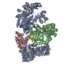
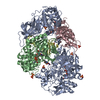
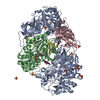
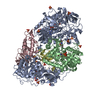
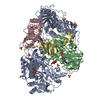
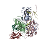
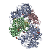
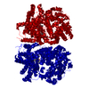
| Details | The biological assembly is a heterotetramer. |
-Components
-Heterotetrameric sarcosine oxidase ... , 4 types, 4 molecules ABCD
| #1: Protein | Mass: 103218.539 Da / Num. of mol.: 1 Source method: isolated from a genetically manipulated source Source: (gene. exp.) Stenotrophomonas maltophilia (bacteria)Gene: soxA / Plasmid: pET28b / Species (production host): Escherichia coli / Production host: |
|---|---|
| #2: Protein | Mass: 44117.762 Da / Num. of mol.: 1 Source method: isolated from a genetically manipulated source Source: (gene. exp.) Stenotrophomonas maltophilia (bacteria)Gene: soxB / Plasmid: pET28b / Species (production host): Escherichia coli / Production host: |
| #3: Protein | Mass: 22235.105 Da / Num. of mol.: 1 Source method: isolated from a genetically manipulated source Source: (gene. exp.) Stenotrophomonas maltophilia (bacteria)Gene: soxG / Plasmid: pET28b / Species (production host): Escherichia coli / Production host: |
| #4: Protein | Mass: 11395.863 Da / Num. of mol.: 1 Source method: isolated from a genetically manipulated source Source: (gene. exp.) Stenotrophomonas maltophilia (bacteria)Gene: soxD / Plasmid: pET28b / Species (production host): Escherichia coli / Production host: |
-Non-polymers , 8 types, 1487 molecules 


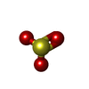


| #5: Chemical |  Mass: 112.083 Da / Num. of mol.: 2 / Source method: obtained synthetically / Formula: C5H4O3 Mass: 112.083 Da / Num. of mol.: 2 / Source method: obtained synthetically / Formula: C5H4O3#6: Chemical | ChemComp-NAD / |  Mass: 663.425 Da / Num. of mol.: 1 / Source method: obtained synthetically / Formula: C21H27N7O14P2 / Comment: NAD*YM Mass: 663.425 Da / Num. of mol.: 1 / Source method: obtained synthetically / Formula: C21H27N7O14P2 / Comment: NAD*YM#7: Chemical | ChemComp-GOL / |  Mass: 92.094 Da / Num. of mol.: 1 / Source method: obtained synthetically / Formula: C3H8O3 Mass: 92.094 Da / Num. of mol.: 1 / Source method: obtained synthetically / Formula: C3H8O3#8: Chemical | ChemComp-SO3 / |  Mass: 80.063 Da / Num. of mol.: 1 / Source method: obtained synthetically / Formula: SO3 Mass: 80.063 Da / Num. of mol.: 1 / Source method: obtained synthetically / Formula: SO3#9: Chemical | ChemComp-FAD / |  Mass: 785.550 Da / Num. of mol.: 1 / Source method: obtained synthetically / Formula: C27H33N9O15P2 / Comment: FAD*YM Mass: 785.550 Da / Num. of mol.: 1 / Source method: obtained synthetically / Formula: C27H33N9O15P2 / Comment: FAD*YM#10: Chemical | ChemComp-FMN / |  Mass: 456.344 Da / Num. of mol.: 1 / Source method: obtained synthetically / Formula: C17H21N4O9P Mass: 456.344 Da / Num. of mol.: 1 / Source method: obtained synthetically / Formula: C17H21N4O9P#11: Chemical | ChemComp-ZN / |  Mass: 65.409 Da / Num. of mol.: 1 / Source method: obtained synthetically / Formula: Zn Mass: 65.409 Da / Num. of mol.: 1 / Source method: obtained synthetically / Formula: Zn#12: Water | ChemComp-HOH / | Mass: 18.015 Da / Num. of mol.: 1479 / Source method: isolated from a natural source / Formula: H2O |
|---|
-Details
| Has protein modification | Y |
|---|
-Experimental details
-Experiment
| Experiment | Method: X-RAY DIFFRACTION / Number of used crystals: 1 |
|---|
- Sample preparation
| Crystal | Density Matthews: 2.64 Å3/Da / Density % sol: 53.44 % |
|---|---|
| Crystal grow | Temperature: 296 K / Method: vapor diffusion, hanging drop / pH: 8.5 Details: 17% PEG 20000, 0.1M Tris, 0.04M sodium fuorate and 0.01M sodium sulfite., pH 8.5, VAPOR DIFFUSION, HANGING DROP, temperature 296K |
-Data collection
| Diffraction | Mean temperature: 100 K |
|---|---|
| Diffraction source | Source: SYNCHROTRON / Site: APS  / Beamline: 14-BM-C / Wavelength: 0.9 Å / Beamline: 14-BM-C / Wavelength: 0.9 Å |
| Detector | Type: ADSC QUANTUM 315 / Detector: CCD / Date: Apr 2, 2004 |
| Radiation | Monochromator: APS BEAMLINE 14 BMC / Protocol: SINGLE WAVELENGTH / Monochromatic (M) / Laue (L): M / Scattering type: x-ray |
| Radiation wavelength | Wavelength: 0.9 Å / Relative weight: 1 |
| Reflection | Resolution: 1.85→40 Å / Num. all: 164316 / Num. obs: 157579 / % possible obs: 95.9 % / Observed criterion σ(F): -3 / Observed criterion σ(I): -3 / Redundancy: 4 % / Biso Wilson estimate: 16.6 Å2 / Rmerge(I) obs: 0.089 / Net I/σ(I): 12.5 |
| Reflection shell | Resolution: 1.85→1.92 Å / Redundancy: 3.6 % / Rmerge(I) obs: 0.325 / Mean I/σ(I) obs: 2.7 / Num. unique all: 14815 / % possible all: 91.1 |
- Processing
| Software |
| |||||||||||||||||||||||||||||||||||||||||||||||||
|---|---|---|---|---|---|---|---|---|---|---|---|---|---|---|---|---|---|---|---|---|---|---|---|---|---|---|---|---|---|---|---|---|---|---|---|---|---|---|---|---|---|---|---|---|---|---|---|---|---|---|
| Refinement | Method to determine structure: MOLECULAR REPLACEMENT Starting model: pdb entry 2GAH Resolution: 1.85→40 Å / Rfactor Rfree error: 0.003 / Data cutoff high absF: 1117856.93 / Data cutoff low absF: 0 / Isotropic thermal model: Isotropic / Cross valid method: THROUGHOUT / σ(F): 0 / σ(I): 0 / Stereochemistry target values: Engh & Huber
| |||||||||||||||||||||||||||||||||||||||||||||||||
| Displacement parameters | Biso mean: 30.1 Å2 | |||||||||||||||||||||||||||||||||||||||||||||||||
| Refine analyze |
| |||||||||||||||||||||||||||||||||||||||||||||||||
| Refinement step | Cycle: LAST / Resolution: 1.85→40 Å
| |||||||||||||||||||||||||||||||||||||||||||||||||
| Refine LS restraints |
| |||||||||||||||||||||||||||||||||||||||||||||||||
| LS refinement shell | Refine-ID: X-RAY DIFFRACTION / Total num. of bins used: 6
|
