 Movie
Movie Controller


+ Open data

- Basic information
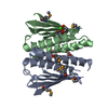
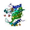
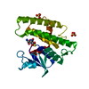
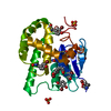
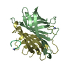
| Entry | Database: PDB / ID: 2g0j | |||||||||
|---|---|---|---|---|---|---|---|---|---|---|
| Title | Crystal structure of SMU.848 from Streptococcus mutans | |||||||||
 Components Components | hypothetical protein SMU.848 | |||||||||
 Keywords Keywords | UNKNOWN FUNCTION / 2-layer (alpha-beta)-sandwich | |||||||||
| Function / homology |  Function and homology information Function and homology informationcysteine-type peptidase activity / ribosome biogenesis / proteolysis / metal ion binding Similarity search - Function | |||||||||
| Biological species |  Streptococcus mutans (bacteria) Streptococcus mutans (bacteria) | |||||||||
| Method |  X-RAY DIFFRACTION / SYNCHROTRON / MOLECULAR REPLACEMENT / Resolution: 2.8 Å X-RAY DIFFRACTION / SYNCHROTRON / MOLECULAR REPLACEMENT / Resolution: 2.8 Å | |||||||||
 Authors Authors | Hou, H.-F. / Gao, Z.-Q. / Li, L.-F. / Liang, Y.-H. / Su, X.-D. / Dong, Y.-H. | |||||||||
 Citation Citation | Journal: To be Published Title: Crystal structure of SMU.848 from Streptococcus mutans Authors: Hou, H.-F. / Gao, Z.-Q. / Xu, J.-H. / Xu, R. / Li, L.-Q. / Li, L.-F. / Liang, Y.-H. / Su, X.-D. / Liu, P. / Xian, D.-C. / Dong, Y.-H. | |||||||||
| History |
|
- Structure visualization
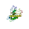
| Structure viewer | Molecule: MolmilJmol/JSmol |
|---|
- Downloads & links
-Download
| PDBx/mmCIF format | 2g0j.cif.gz | 98.7 KB | Display | PDBx/mmCIF format |
|---|---|---|---|---|
| PDB format | pdb2g0j.ent.gz | 75.7 KB | Display | PDB format |
| PDBx/mmJSON format | 2g0j.json.gz | Tree view | PDBx/mmJSON format | |
| Others |  Other downloads Other downloads |
-Validation report
| Arichive directory | https://data.pdbj.org/pub/pdb/validation_reports/g0/2g0jftp://data.pdbj.org/pub/pdb/validation_reports/g0/2g0j | HTTPS FTP |
|---|
-Related structure data


-Links
 PDBj
PDBj- Assembly
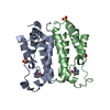
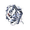
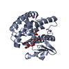
| Deposited unit | 
| ||||||||
|---|---|---|---|---|---|---|---|---|---|
| 1 | 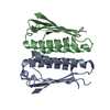
| ||||||||
| 2 | 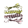
| ||||||||
| 3 | 
| ||||||||
| Unit cell |
|
-Components
| #1: Protein | Mass: 15707.851 Da / Num. of mol.: 4 Source method: isolated from a genetically manipulated source Source: (gene. exp.) Streptococcus mutans (bacteria) / Plasmid: Pet28(a) / Species (production host): Escherichia coli / Production host: #2: Water | ChemComp-HOH / |  Mass: 18.015 Da / Num. of mol.: 74 / Source method: isolated from a natural source / Formula: H2O Mass: 18.015 Da / Num. of mol.: 74 / Source method: isolated from a natural source / Formula: H2O |
|---|
-Experimental details
-Experiment
| Experiment | Method: X-RAY DIFFRACTION / Number of used crystals: 1 |
|---|
- Sample preparation
| Crystal | Density Matthews: 1.83 Å3/Da / Density % sol: 32.65 % |
|---|---|
| Crystal grow | Temperature: 289 K / Method: vapor diffusion, hanging drop / pH: 7.5 Details: 0.1M HEPES-NaOH, PH 7.5, 28% PEG 400, 0.2M CaCl2, VAPOR DIFFUSION, HANGING DROP, temperature 289K |
-Data collection
| Diffraction | Mean temperature: 100 K | |||||||||||||||||||||||||||||||||||||||||||||||||||||||||||||||||||||||||||||
|---|---|---|---|---|---|---|---|---|---|---|---|---|---|---|---|---|---|---|---|---|---|---|---|---|---|---|---|---|---|---|---|---|---|---|---|---|---|---|---|---|---|---|---|---|---|---|---|---|---|---|---|---|---|---|---|---|---|---|---|---|---|---|---|---|---|---|---|---|---|---|---|---|---|---|---|---|---|---|
| Diffraction source | Source: SYNCHROTRON / Site: BSRF  / Beamline: 3W1A / Wavelength: 1 Å / Beamline: 3W1A / Wavelength: 1 Å | |||||||||||||||||||||||||||||||||||||||||||||||||||||||||||||||||||||||||||||
| Detector | Type: MARRESEARCH / Detector: CCD / Date: Feb 20, 2005 | |||||||||||||||||||||||||||||||||||||||||||||||||||||||||||||||||||||||||||||
| Radiation | Monochromator: Si(111) double-crystal / Protocol: SINGLE WAVELENGTH / Monochromatic (M) / Laue (L): M / Scattering type: x-ray | |||||||||||||||||||||||||||||||||||||||||||||||||||||||||||||||||||||||||||||
| Radiation wavelength | Wavelength: 1 Å / Relative weight: 1 | |||||||||||||||||||||||||||||||||||||||||||||||||||||||||||||||||||||||||||||
| Reflection | Redundancy: 6 % / Av σ(I) over netI: 7 / Number: 71233 / Rmerge(I) obs: 0.11 / Χ2: 1.06 / D res high: 2.8 Å / D res low: 50 Å / Num. obs: 11810 / % possible obs: 99.7 | |||||||||||||||||||||||||||||||||||||||||||||||||||||||||||||||||||||||||||||
| Diffraction reflection shell | ID: 1
| |||||||||||||||||||||||||||||||||||||||||||||||||||||||||||||||||||||||||||||
| Reflection | Resolution: 2.8→50 Å / Num. all: 11846 / Num. obs: 11810 / % possible obs: 99.7 % / Observed criterion σ(F): 1 / Observed criterion σ(I): 1 / Redundancy: 6 % / Rmerge(I) obs: 0.11 / Χ2: 1.062 / Net I/σ(I): 7 | |||||||||||||||||||||||||||||||||||||||||||||||||||||||||||||||||||||||||||||
| Reflection shell | Resolution: 2.8→2.9 Å / % possible obs: 99.7 % / Redundancy: 6 % / Rmerge(I) obs: 0.599 / Num. unique obs: 1149 / Χ2: 1.001 / % possible all: 99.7 |
- Processing
| Software |
| ||||||||||||||||||||||||||||
|---|---|---|---|---|---|---|---|---|---|---|---|---|---|---|---|---|---|---|---|---|---|---|---|---|---|---|---|---|---|
| Refinement | Method to determine structure: MOLECULAR REPLACEMENT / Resolution: 2.8→50 Å / σ(F): 0 / Stereochemistry target values: Engh & Huber
| ||||||||||||||||||||||||||||
| Solvent computation | Bsol: 41.63 Å2 | ||||||||||||||||||||||||||||
| Displacement parameters | Biso mean: 38.197 Å2
| ||||||||||||||||||||||||||||
| Refinement step | Cycle: LAST / Resolution: 2.8→50 Å
| ||||||||||||||||||||||||||||
| Xplor file |
|
