 Movie
Movie Controller

[English] 日本語
 Yorodumi
- PDB-2fug: Crystal structure of the hydrophilic domain of respiratory comple... -
+ Open data

- Basic information
| Entry | Database: PDB / ID: 2fug | ||||||
|---|---|---|---|---|---|---|---|
| Title | Crystal structure of the hydrophilic domain of respiratory complex I from Thermus thermophilus | ||||||
 Components Components |
| ||||||
 Keywords Keywords | OXIDOREDUCTASE / ELECTRON TRANSPORT / RESPIRATORY CHAIN | ||||||
| Function / homology |  Function and homology information Function and homology informationTranslocases; Catalysing the translocation of protons; Linked to oxidoreductase reactions / NADH dehydrogenase (quinone) (non-electrogenic) activity / molybdopterin cofactor binding / NADH dehydrogenase activity / iron-sulfur cluster assembly / electron transport coupled proton transport / respiratory chain complex I / NADH dehydrogenase (ubiquinone) activity / quinone binding / ATP synthesis coupled electron transport ...Translocases; Catalysing the translocation of protons; Linked to oxidoreductase reactions / NADH dehydrogenase (quinone) (non-electrogenic) activity / molybdopterin cofactor binding / NADH dehydrogenase activity / iron-sulfur cluster assembly / electron transport coupled proton transport / respiratory chain complex I / NADH dehydrogenase (ubiquinone) activity / quinone binding / ATP synthesis coupled electron transport / ferric iron binding / aerobic respiration / 2 iron, 2 sulfur cluster binding / NAD binding / FMN binding / 4 iron, 4 sulfur cluster binding / iron ion binding / metal ion binding / plasma membrane Similarity search - Function | ||||||
| Biological species |   Thermus thermophilus (bacteria) Thermus thermophilus (bacteria) | ||||||
| Method |  X-RAY DIFFRACTION / SYNCHROTRON / MIRAS / Resolution: 3.3 Å X-RAY DIFFRACTION / SYNCHROTRON / MIRAS / Resolution: 3.3 Å | ||||||
 Authors Authors | Sazanov, L.A. / Hinchliffe, P. | ||||||
 Citation Citation | Journal: Science / Year: 2006 Title: Structure of the hydrophilic domain of respiratory complex I from Thermus thermophilus. Authors: Sazanov, L.A. / Hinchliffe, P. #1: Journal: Science / Year: 2005Title: Organization of iron-sulfur clusters in respiratory complex I Authors: Hinchliffe, P. / Sazanov, L.A. | ||||||
| History |
|
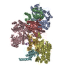
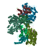
- Structure visualization
| Structure viewer | Molecule: MolmilJmol/JSmol |
|---|
- Downloads & links
-Download
| PDBx/mmCIF format | 2fug.cif.gz | 1.8 MB | Display | PDBx/mmCIF format |
|---|---|---|---|---|
| PDB format | pdb2fug.ent.gz | 1.5 MB | Display | PDB format |
| PDBx/mmJSON format | 2fug.json.gz | Tree view | PDBx/mmJSON format | |
| Others |  Other downloads Other downloads |
-Validation report
| Summary document | 2fug_validation.pdf.gz | 1.7 MB | Display | wwPDB validaton report |
|---|---|---|---|---|
| Full document | 2fug_full_validation.pdf.gz | 2.3 MB | Display | |
| Data in XML | 2fug_validation.xml.gz | 427.5 KB | Display | |
| Data in CIF | 2fug_validation.cif.gz | 551.3 KB | Display | |
| Arichive directory | https://data.pdbj.org/pub/pdb/validation_reports/fu/2fugftp://data.pdbj.org/pub/pdb/validation_reports/fu/2fug | HTTPS FTP |
-Related structure data
| Similar structure data |
|---|


-Links
 PDBj
PDBj


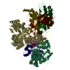
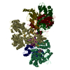
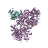
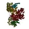
- Assembly
| Deposited unit | 
| ||||||||
|---|---|---|---|---|---|---|---|---|---|
| 1 | 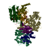
| ||||||||
| 2 | 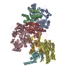
| ||||||||
| 3 | 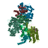
| ||||||||
| 4 | 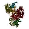
| ||||||||
| Unit cell |
|
-Components
-NADH-quinone oxidoreductase chain ... , 7 types, 28 molecules 1AJS2BKT3CLU4DMV5ENW6FOX9GPY
| #1: Protein | Mass: 48693.715 Da / Num. of mol.: 4 / Fragment: Hydrophilic domain / Source method: isolated from a natural source / Source: (natural) Thermus thermophilus (bacteria) / Strain: HB8 / References: UniProt: Q56222, NADH dehydrogenase (quinone)#2: Protein | Mass: 20309.162 Da / Num. of mol.: 4 / Source method: isolated from a natural source / Source: (natural) Thermus thermophilus (bacteria) / Strain: HB8 / References: UniProt: Q56221, NADH dehydrogenase (quinone)#3: Protein | Mass: 86656.203 Da / Num. of mol.: 4 / Source method: isolated from a natural source / Source: (natural) Thermus thermophilus (bacteria) / Strain: HB8 / References: UniProt: Q56223, NADH dehydrogenase (quinone)#4: Protein | Mass: 46428.027 Da / Num. of mol.: 4 / Source method: isolated from a natural source / Source: (natural) Thermus thermophilus (bacteria) / Strain: HB8 / References: UniProt: Q56220, NADH dehydrogenase (quinone)#5: Protein | Mass: 23893.254 Da / Num. of mol.: 4 / Source method: isolated from a natural source / Source: (natural) Thermus thermophilus (bacteria) / Strain: HB8 / References: UniProt: Q56219, NADH dehydrogenase (quinone)#6: Protein | Mass: 20262.564 Da / Num. of mol.: 4 / Source method: isolated from a natural source / Source: (natural) Thermus thermophilus (bacteria) / Strain: HB8 / References: UniProt: Q56218, NADH dehydrogenase (quinone)#7: Protein | Mass: 20106.309 Da / Num. of mol.: 4 / Source method: isolated from a natural source / Source: (natural) Thermus thermophilus (bacteria) / Strain: HB8 / References: UniProt: Q56224, NADH dehydrogenase (quinone) |
|---|
-Protein , 1 types, 4 molecules 7HQZ
| #8: Protein | Mass: 14812.074 Da / Num. of mol.: 4 / Source method: isolated from a natural source / Source: (natural) Thermus thermophilus (bacteria) / Strain: HB8 / References: GenBank: 55771878, UniProt: Q5SKZ7*PLUS |
|---|
-Non-polymers , 3 types, 40 molecules 


| #9: Chemical | ChemComp-SF4 /  Mass: 351.640 Da / Num. of mol.: 28 / Source method: obtained synthetically / Formula: Fe4S4 Mass: 351.640 Da / Num. of mol.: 28 / Source method: obtained synthetically / Formula: Fe4S4#10: Chemical | ChemComp-FES /  Mass: 175.820 Da / Num. of mol.: 8 / Source method: obtained synthetically / Formula: Fe2S2 Mass: 175.820 Da / Num. of mol.: 8 / Source method: obtained synthetically / Formula: Fe2S2#11: Chemical | ChemComp-FMN /  Mass: 456.344 Da / Num. of mol.: 4 / Source method: obtained synthetically / Formula: C17H21N4O9P Mass: 456.344 Da / Num. of mol.: 4 / Source method: obtained synthetically / Formula: C17H21N4O9P |
|---|
-Details
| Has protein modification | Y |
|---|
-Experimental details
-Experiment
| Experiment | Method: X-RAY DIFFRACTION / Number of used crystals: 1 |
|---|
- Sample preparation
| Crystal | Density Matthews: 3.12 Å3/Da / Density % sol: 60.52 % |
|---|---|
| Crystal grow | Temperature: 295 K / Method: vapor diffusion, sitting drop / pH: 7.5 Details: 0.1M HEPES pH 7.5, 0.4-0.5M NaCl, 0.1M CaCl2 and 8-10% PEG4000, VAPOR DIFFUSION, SITTING DROP, temperature 295K |
-Data collection
| Diffraction | Mean temperature: 100 K |
|---|---|
| Diffraction source | Source: SYNCHROTRON / Site: ESRF  / Beamline: ID29 / Wavelength: 0.97564 Å / Beamline: ID29 / Wavelength: 0.97564 Å |
| Detector | Type: ADSC QUANTUM 210 / Detector: CCD / Date: Nov 7, 2004 |
| Radiation | Monochromator: Si(311) / Protocol: SINGLE WAVELENGTH / Monochromatic (M) / Laue (L): M / Scattering type: x-ray |
| Radiation wavelength | Wavelength: 0.97564 Å / Relative weight: 1 |
| Reflection | Resolution: 3.3→29.988 Å / Num. all: 196320 / Num. obs: 195669 / % possible obs: 92.7 % / Observed criterion σ(F): 0 / Observed criterion σ(I): 0 / Redundancy: 3.2 % / Biso Wilson estimate: 47.5 Å2 / Rmerge(I) obs: 0.158 / Rsym value: 0.158 / Net I/σ(I): 6.1 |
| Reflection shell | Resolution: 3.3→3.48 Å / % possible obs: 89.2 % / Redundancy: 3.2 % / Rmerge(I) obs: 0.552 / Mean I/σ(I) obs: 1.7 / Num. measured all: 84269 / Num. unique all: 26742 / Num. unique obs: 26742 / Rsym value: 0.552 / % possible all: 89.2 |
- Processing
| Software |
| ||||||||||||||||||||||||||||
|---|---|---|---|---|---|---|---|---|---|---|---|---|---|---|---|---|---|---|---|---|---|---|---|---|---|---|---|---|---|
| Refinement | Method to determine structure: MIRAS / Resolution: 3.3→20 Å / Rfactor Rfree error: 0.005 / FOM work R set: 0.744 / Isotropic thermal model: isotropic restrained / Cross valid method: THROUGHOUT / σ(F): 0 / σ(I): 0 / Stereochemistry target values: Engh & Huber Details: The number of reflections used in refinement included two derivatives and Fe-edge, at lower resolution, were merged in SHARP during phasing.
| ||||||||||||||||||||||||||||
| Displacement parameters | Biso mean: 64.8 Å2 | ||||||||||||||||||||||||||||
| Refine analyze |
| ||||||||||||||||||||||||||||
| Refinement step | Cycle: LAST / Resolution: 3.3→20 Å
| ||||||||||||||||||||||||||||
| Refine LS restraints |
| ||||||||||||||||||||||||||||
| LS refinement shell | Resolution: 3.3→3.42 Å / Rfactor Rfree error: 0.021 / Total num. of bins used: 10
|
