 Movie
Movie Controller


+ Open data

- Basic information
| Entry | Database: PDB / ID: 2ftk | ||||||
|---|---|---|---|---|---|---|---|
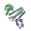
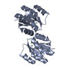
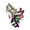
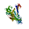
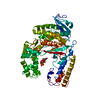
| Title | berylloflouride Spo0F complex with Spo0B | ||||||
 Components Components |
| ||||||
 Keywords Keywords | TRANSFERASE / Sporulation / Spo0F / Spo0B phosphorelay | ||||||
| Function / homology |  Function and homology information Function and homology informationTransferases; Transferring phosphorus-containing groups / phosphorelay response regulator activity / sporulation resulting in formation of a cellular spore / phosphorelay sensor kinase activity / phosphorelay signal transduction system / kinase activity / metal ion binding / cytoplasm Similarity search - Function | ||||||
| Biological species |  | ||||||
| Method |  X-RAY DIFFRACTION / SYNCHROTRON / MOLECULAR REPLACEMENT / Resolution: 3.05 Å X-RAY DIFFRACTION / SYNCHROTRON / MOLECULAR REPLACEMENT / Resolution: 3.05 Å | ||||||
 Authors Authors | Varughese, K.I. | ||||||
 Citation Citation | Journal: J.Bacteriol. / Year: 2006 Title: The Crystal Structure of Beryllofluoride Spo0F in Complex with the Phosphotransferase Spo0B Represents a Phosphotransfer Pretransition State. Authors: Varughese, K.I. / Tsigelny, I. / Zhao, H. | ||||||
| History |
|
- Structure visualization
| Structure viewer | Molecule: MolmilJmol/JSmol |
|---|
- Downloads & links
-Download
| PDBx/mmCIF format | 2ftk.cif.gz | 252.2 KB | Display | PDBx/mmCIF format |
|---|---|---|---|---|
| PDB format | pdb2ftk.ent.gz | 202.5 KB | Display | PDB format |
| PDBx/mmJSON format | 2ftk.json.gz | Tree view | PDBx/mmJSON format | |
| Others |  Other downloads Other downloads |
-Validation report
| Arichive directory | https://data.pdbj.org/pub/pdb/validation_reports/ft/2ftkftp://data.pdbj.org/pub/pdb/validation_reports/ft/2ftk | HTTPS FTP |
|---|
-Related structure data
| Related structure data | 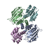 1f51S S: Starting model for refinement |
|---|---|
| Similar structure data |


-Links
 PDBj
PDBj

- Assembly
| Deposited unit | 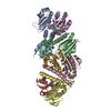
| ||||||||
|---|---|---|---|---|---|---|---|---|---|
| 1 | 
| ||||||||
| 2 | 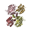
| ||||||||
| Unit cell |
|
-Components
| #1: Protein | Mass: 22573.559 Da / Num. of mol.: 4 Source method: isolated from a genetically manipulated source Source: (gene. exp.) References: UniProt: P06535, Transferases; Transferring phosphorus-containing groups #2: Protein | Mass: 14233.564 Da / Num. of mol.: 4 / Mutation: Y13S Source method: isolated from a genetically manipulated source Source: (gene. exp.) References: UniProt: P06628, Transferases; Transferring phosphorus-containing groups #3: Chemical | ChemComp-MG /   Mass: 24.305 Da / Num. of mol.: 4 / Source method: obtained synthetically / Formula: Mg Mass: 24.305 Da / Num. of mol.: 4 / Source method: obtained synthetically / Formula: MgHas protein modification | Y | |
|---|
-Experimental details
-Experiment
| Experiment | Method: X-RAY DIFFRACTION / Number of used crystals: 1 |
|---|
- Sample preparation
| Crystal | Density Matthews: 2.48 Å3/Da / Density % sol: 50.46 % |
|---|---|
| Crystal grow | Temperature: 297 K / Method: vapor diffusion / pH: 8.1 Details: 0.5 M KCl, 25% PEG2000, 100mM Tris HCl ph8.1, VAPOR DIFFUSION, temperature 297K |
-Data collection
| Diffraction | Mean temperature: 90 K |
|---|---|
| Diffraction source | Source: SYNCHROTRON / Site: SSRL  / Beamline: BL9-1 / Beamline: BL9-1 |
| Detector | Type: ADSC QUANTUM 315 / Detector: CCD / Date: Jul 14, 2005 |
| Radiation | Monochromator: Flat mirror and bent monochromator / Protocol: SINGLE WAVELENGTH / Monochromatic (M) / Laue (L): M / Scattering type: x-ray |
| Radiation wavelength | Relative weight: 1 |
| Reflection | Resolution: 3.05→19.86 Å / Num. all: 28073 / Num. obs: 28073 / % possible obs: 98.6 % / Observed criterion σ(F): 0 / Observed criterion σ(I): 0 |
| Reflection shell | Resolution: 3.05→3.24 Å / % possible all: 90.8 |
- Processing
| Software |
| |||||||||||||||||||||||||
|---|---|---|---|---|---|---|---|---|---|---|---|---|---|---|---|---|---|---|---|---|---|---|---|---|---|---|
| Refinement | Method to determine structure: MOLECULAR REPLACEMENT Starting model: 1F51 Resolution: 3.05→19.86 Å / Cross valid method: R free / σ(F): 0 / σ(I): 0 / Stereochemistry target values: Engh & Huber
| |||||||||||||||||||||||||
| Refine analyze |
| |||||||||||||||||||||||||
| Refinement step | Cycle: LAST / Resolution: 3.05→19.86 Å
| |||||||||||||||||||||||||
| Refine LS restraints |
|
