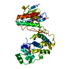
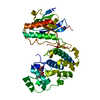
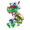
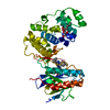
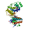
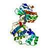
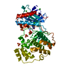
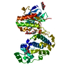
Entry Database : PDB / ID : 2fstTitle mitogen activated protein kinase p38alpha (D176A+F327L) activating mutant Mitogen-activated protein kinase 14 Keywords / / / / / / Function / homology Function Domain/homology Component
/ / / / / / / / / / / / / / / / / / / / / / / / / / / / / / / / / / / / / / / / / / / / / / / / / / / / / / / / / / / / / / / / / / / / / / / / / / / / / / / / / / / / / / / / / / / / / / / / / / / / / / / / / / / / / / / / / / / / / / / / / / / / / / / / / / / / / / / / Biological species Homo sapiens (human)Method / / / Resolution : 1.45 Å Authors Diskin, R. / Livnah, O. Journal : J.Mol.Biol. / Year : 2007Title : Structures of p38alpha Active Mutants Reveal Conformational Changes in L16 Loop that Induce Autophosphorylation and ActivationAuthors : Diskin, R. / Lebendiker, M. / Engelberg, D. / Livnah, O. History Deposition Jan 23, 2006 Deposition site / Processing site Revision 1.0 Dec 5, 2006 Provider / Type Revision 1.1 May 1, 2008 Group Revision 1.2 Jul 13, 2011 Group Revision 1.3 Jul 29, 2020 Group Data collection / Database references ... Data collection / Database references / Derived calculations / Structure summary Category chem_comp / entity ... chem_comp / entity / pdbx_chem_comp_identifier / pdbx_entity_nonpoly / struct_ref_seq_dif / struct_site / struct_site_gen Item _chem_comp.mon_nstd_flag / _chem_comp.name ... _chem_comp.mon_nstd_flag / _chem_comp.name / _chem_comp.type / _entity.pdbx_description / _pdbx_entity_nonpoly.name / _struct_ref_seq_dif.details Description / Provider / Type Revision 1.4 Nov 10, 2021 Group / Structure summary / Category / database_2 / struct_ref_seq_difItem _chem_comp.pdbx_synonyms / _database_2.pdbx_DOI ... _chem_comp.pdbx_synonyms / _database_2.pdbx_DOI / _database_2.pdbx_database_accession / _struct_ref_seq_dif.details Revision 1.5 May 29, 2024 Group / Category / chem_comp_bond
Show all Show less
 Movie
Movie Controller

 Yorodumi
 Open data
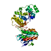
 Basic information
 Components
 Keywords
 Function and homology information
 Homo sapiens (human)
 X-RAY DIFFRACTION /
  Authors
 Citation
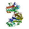
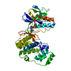
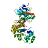
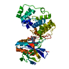
 Structure visualization
 Downloads & links
 Other downloads


 PDBj
PDBj


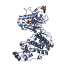
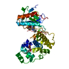
 Assembly
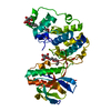


 Type: D-saccharide / Mass: 292.369 Da / Num. of mol.: 3
 Mass: 18.015 Da / Num. of mol.: 435 / Source method: isolated from a natural source / Formula: H2O
 Sample preparation
 / Beamline: ID14-4 / Wavelength: 0.925 Å
 Processing