 Movie
Movie Controller


+ Open data

- Basic information
| Entry | Database: PDB / ID: 2fqz | ||||||
|---|---|---|---|---|---|---|---|
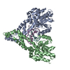
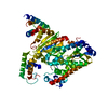
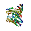
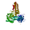
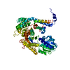
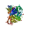
| Title | Metal-depleted Ecl18kI in complex with uncleaved DNA | ||||||
 Components Components |
| ||||||
 Keywords Keywords | Hydrolase/DNA / ECL18KI-DNA COMPLEX / TYPE II RESTRICTION ENDONUCLEASE / NUCLEOTIDE FLIPPING / BASE EXTRUSION / Hydrolase-DNA COMPLEX | ||||||
| Function / homology |  Function and homology information Function and homology informationtype II site-specific deoxyribonuclease activity / DNA restriction-modification system / DNA binding Similarity search - Function | ||||||
| Biological species |  Enterobacter cloacae (bacteria) Enterobacter cloacae (bacteria) | ||||||
| Method |  X-RAY DIFFRACTION / SYNCHROTRON / MAD / Resolution: 2 Å X-RAY DIFFRACTION / SYNCHROTRON / MAD / Resolution: 2 Å | ||||||
 Authors Authors | Bochtler, M. / Szczepanowski, R.H. / Tamulaitis, G. / Grazulis, S. / Czapinska, H. / Manakova, E. / Siksnys, V. | ||||||
 Citation Citation | Journal: Embo J. / Year: 2006 Title: Nucleotide flips determine the specificity of the Ecl18kI restriction endonuclease Authors: Bochtler, M. / Szczepanowski, R.H. / Tamulaitis, G. / Grazulis, S. / Czapinska, H. / Manakova, E. / Siksnys, V. #1: Journal: FEBS Lett. / Year: 2002 Title: Alternative arrangements of catalytic residues at the active sites of restriction enzymes. Authors: Tamulaitis, G. / Solonin, A.S. / Siksnys, V. #2: Journal: FEBS Lett. / Year: 1998 Title: The Ecl18kI restriction-modification system: cloning, expression, properties of the purified enzymes. Authors: Denjmukhametov, M.M. / Brevnov, M.G. / Zakharova, M.V. / Repyk, A.V. / Solonin, A.S. / Petrauskene, O.V. / Gromova, E.S. | ||||||
| History |
| ||||||
| Remark 300 | BIOMOLECULE 1, 2 THIS ENTRY CONTAINS THE CRYSTALLOGRAPHIC ASYMMETRIC UNIT WHICH CONSISTS OF 8 ...BIOMOLECULE 1, 2 THIS ENTRY CONTAINS THE CRYSTALLOGRAPHIC ASYMMETRIC UNIT WHICH CONSISTS OF 8 CHAIN(S). SEE REMARK 350 FOR INFORMATION ON GENERATING THE BIOLOGICAL MOLECULE(S). UNDER PHYSIOLOGICAL CONDITIONS Ecl18KI ENDONUCLEASE EXISTS PREDOMINANTLY AS A DIMER (CHAINS AB OR CD), HOWEVER THERE IS SOME EVIDENCE THAT AT HIGH CONCENTRATIONS AND IN SOME CONDITIONS DIMERS MAY ASSOCIATE. |
- Structure visualization
| Structure viewer | Molecule: MolmilJmol/JSmol |
|---|
- Downloads & links
-Download
| PDBx/mmCIF format | 2fqz.cif.gz | 289.5 KB | Display | PDBx/mmCIF format |
|---|---|---|---|---|
| PDB format | pdb2fqz.ent.gz | 234.8 KB | Display | PDB format |
| PDBx/mmJSON format | 2fqz.json.gz | Tree view | PDBx/mmJSON format | |
| Others |  Other downloads Other downloads |
-Validation report
| Arichive directory | https://data.pdbj.org/pub/pdb/validation_reports/fq/2fqzftp://data.pdbj.org/pub/pdb/validation_reports/fq/2fqz | HTTPS FTP |
|---|
-Related structure data


-Links
 PDBj
PDBj


- Assembly
| Deposited unit | 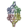
| ||||||||
|---|---|---|---|---|---|---|---|---|---|
| 1 | 
| ||||||||
| 2 | 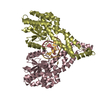
| ||||||||
| Unit cell |
|
-Components
| #1: DNA chain | Mass: 2741.800 Da / Num. of mol.: 2 / Source method: obtained synthetically #2: DNA chain | Mass: 2732.786 Da / Num. of mol.: 2 / Source method: obtained synthetically #3: Protein | Mass: 35938.785 Da / Num. of mol.: 4 Source method: isolated from a genetically manipulated source Source: (gene. exp.) Enterobacter cloacae (bacteria) / Gene: ecl18kIR / Plasmid: pUC129 / Production host: #4: Water | ChemComp-HOH / |  Mass: 18.015 Da / Num. of mol.: 555 / Source method: isolated from a natural source / Formula: H2O Mass: 18.015 Da / Num. of mol.: 555 / Source method: isolated from a natural source / Formula: H2O |
|---|
-Experimental details
-Experiment
| Experiment | Method: X-RAY DIFFRACTION / Number of used crystals: 3 |
|---|
- Sample preparation
| Crystal | Density Matthews: 2.26 Å3/Da / Density % sol: 45.64 % Description: Data for refinement were collected at 0.8019 A. MAD data were collected on a bromide soaked crystal (1.05, 0.900, 0.9198, 0.9200) and on a crystal of the selenomethionine variant (0.9792, 0.9795) |
|---|---|
| Crystal grow | Temperature: 294 K / Method: vapor diffusion, sitting drop / pH: 4.1 Details: 0.4M (NH4)H2PO4, pH 4.1, VAPOR DIFFUSION, SITTING DROP, temperature 294K |
-Data collection
| Diffraction |
| ||||||||||||||||||||||||
|---|---|---|---|---|---|---|---|---|---|---|---|---|---|---|---|---|---|---|---|---|---|---|---|---|---|
| Diffraction source |
| ||||||||||||||||||||||||
| Detector |
| ||||||||||||||||||||||||
| Radiation |
| ||||||||||||||||||||||||
| Radiation wavelength |
| ||||||||||||||||||||||||
| Reflection | Resolution: 2→20 Å / Num. all: 93417 / Num. obs: 93417 / % possible obs: 93.4 % / Redundancy: 3.6 % / Biso Wilson estimate: 35 Å2 / Rmerge(I) obs: 0.034 / Rsym value: 0.034 / Net I/σ(I): 41 | ||||||||||||||||||||||||
| Reflection shell | Resolution: 2→2.03 Å / Rmerge(I) obs: 0.24 / Mean I/σ(I) obs: 5.12 / Num. unique all: 4852 / Rsym value: 0.24 / % possible all: 97.5 |

- Processing
| Software |
| |||||||||||||||||||||||||||||||||||||||||||||||||||||||||||||||||||||||||||||||||||||||||||||||||||||||||||||||||||||||||||||||||||||||||||||||||||||||||||||||||||||||||||||||||||||||||||||||||||||||||||||||||||||||||||||||||||||||||||||||||||||||||||||||||||||||||||||||||||||||||||||||||||||||||||||||||||||||||||||||||||||
|---|---|---|---|---|---|---|---|---|---|---|---|---|---|---|---|---|---|---|---|---|---|---|---|---|---|---|---|---|---|---|---|---|---|---|---|---|---|---|---|---|---|---|---|---|---|---|---|---|---|---|---|---|---|---|---|---|---|---|---|---|---|---|---|---|---|---|---|---|---|---|---|---|---|---|---|---|---|---|---|---|---|---|---|---|---|---|---|---|---|---|---|---|---|---|---|---|---|---|---|---|---|---|---|---|---|---|---|---|---|---|---|---|---|---|---|---|---|---|---|---|---|---|---|---|---|---|---|---|---|---|---|---|---|---|---|---|---|---|---|---|---|---|---|---|---|---|---|---|---|---|---|---|---|---|---|---|---|---|---|---|---|---|---|---|---|---|---|---|---|---|---|---|---|---|---|---|---|---|---|---|---|---|---|---|---|---|---|---|---|---|---|---|---|---|---|---|---|---|---|---|---|---|---|---|---|---|---|---|---|---|---|---|---|---|---|---|---|---|---|---|---|---|---|---|---|---|---|---|---|---|---|---|---|---|---|---|---|---|---|---|---|---|---|---|---|---|---|---|---|---|---|---|---|---|---|---|---|---|---|---|---|---|---|---|---|---|---|---|---|---|---|---|---|---|---|---|---|---|---|---|---|---|---|---|---|---|---|---|---|---|---|---|---|---|---|---|---|---|---|---|---|---|---|---|---|---|---|---|---|---|---|---|---|---|---|---|---|---|---|---|---|---|---|---|---|---|
| Refinement | Method to determine structure: MAD / Resolution: 2→20 Å / Cor.coef. Fo:Fc: 0.944 / Cor.coef. Fo:Fc free: 0.921 / SU B: 9.219 / SU ML: 0.135 / TLS residual ADP flag: LIKELY RESIDUAL / Isotropic thermal model: TLS / Cross valid method: THROUGHOUT / ESU R: 0.225 / ESU R Free: 0.188 / Stereochemistry target values: MAXIMUM LIKELIHOOD / Details: TLS refinement used
| |||||||||||||||||||||||||||||||||||||||||||||||||||||||||||||||||||||||||||||||||||||||||||||||||||||||||||||||||||||||||||||||||||||||||||||||||||||||||||||||||||||||||||||||||||||||||||||||||||||||||||||||||||||||||||||||||||||||||||||||||||||||||||||||||||||||||||||||||||||||||||||||||||||||||||||||||||||||||||||||||||||
| Solvent computation | Ion probe radii: 0.8 Å / Shrinkage radii: 0.8 Å / VDW probe radii: 1.2 Å / Solvent model: MASK | |||||||||||||||||||||||||||||||||||||||||||||||||||||||||||||||||||||||||||||||||||||||||||||||||||||||||||||||||||||||||||||||||||||||||||||||||||||||||||||||||||||||||||||||||||||||||||||||||||||||||||||||||||||||||||||||||||||||||||||||||||||||||||||||||||||||||||||||||||||||||||||||||||||||||||||||||||||||||||||||||||||
| Displacement parameters | Biso mean: 36.6 Å2
| |||||||||||||||||||||||||||||||||||||||||||||||||||||||||||||||||||||||||||||||||||||||||||||||||||||||||||||||||||||||||||||||||||||||||||||||||||||||||||||||||||||||||||||||||||||||||||||||||||||||||||||||||||||||||||||||||||||||||||||||||||||||||||||||||||||||||||||||||||||||||||||||||||||||||||||||||||||||||||||||||||||
| Refinement step | Cycle: LAST / Resolution: 2→20 Å
| |||||||||||||||||||||||||||||||||||||||||||||||||||||||||||||||||||||||||||||||||||||||||||||||||||||||||||||||||||||||||||||||||||||||||||||||||||||||||||||||||||||||||||||||||||||||||||||||||||||||||||||||||||||||||||||||||||||||||||||||||||||||||||||||||||||||||||||||||||||||||||||||||||||||||||||||||||||||||||||||||||||
| Refine LS restraints |
| |||||||||||||||||||||||||||||||||||||||||||||||||||||||||||||||||||||||||||||||||||||||||||||||||||||||||||||||||||||||||||||||||||||||||||||||||||||||||||||||||||||||||||||||||||||||||||||||||||||||||||||||||||||||||||||||||||||||||||||||||||||||||||||||||||||||||||||||||||||||||||||||||||||||||||||||||||||||||||||||||||||
| LS refinement shell | Resolution: 2→2.03 Å / Total num. of bins used: 20
| |||||||||||||||||||||||||||||||||||||||||||||||||||||||||||||||||||||||||||||||||||||||||||||||||||||||||||||||||||||||||||||||||||||||||||||||||||||||||||||||||||||||||||||||||||||||||||||||||||||||||||||||||||||||||||||||||||||||||||||||||||||||||||||||||||||||||||||||||||||||||||||||||||||||||||||||||||||||||||||||||||||
| Refinement TLS params. | Method: refined / Refine-ID: X-RAY DIFFRACTION
| |||||||||||||||||||||||||||||||||||||||||||||||||||||||||||||||||||||||||||||||||||||||||||||||||||||||||||||||||||||||||||||||||||||||||||||||||||||||||||||||||||||||||||||||||||||||||||||||||||||||||||||||||||||||||||||||||||||||||||||||||||||||||||||||||||||||||||||||||||||||||||||||||||||||||||||||||||||||||||||||||||||
| Refinement TLS group |
|
