 Movie
Movie Controller

[English] 日本語
 Yorodumi
- PDB-2fpm: RadA recombinase in complex with AMP-PNP and high concentration of K+ -
+ Open data

- Basic information
| Entry | Database: PDB / ID: 2fpm | |||||||||
|---|---|---|---|---|---|---|---|---|---|---|
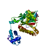
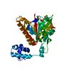
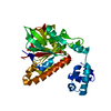
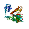
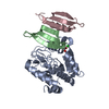
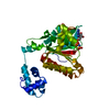
| Title | RadA recombinase in complex with AMP-PNP and high concentration of K+ | |||||||||
 Components Components | DNA repair and recombination protein radA | |||||||||
 Keywords Keywords | RECOMBINATION / ATPASE / PROTEIN-ATP COMPLEX / RADA-ADP COMPLEX / CO-FACTORS / POTASSIUM-DEPENDENCE | |||||||||
| Function / homology |  Function and homology information Function and homology informationATP-dependent DNA damage sensor activity / DNA recombination / damaged DNA binding / DNA repair / ATP binding / identical protein binding Similarity search - Function | |||||||||
| Biological species |  Methanococcus voltae (archaea) Methanococcus voltae (archaea) | |||||||||
| Method |  X-RAY DIFFRACTION / MOLECULAR REPLACEMENT / Resolution: 2 Å X-RAY DIFFRACTION / MOLECULAR REPLACEMENT / Resolution: 2 Å | |||||||||
 Authors Authors | Wu, Y. / Qian, X. / He, Y. / Moya, I.A. / Luo, Y. | |||||||||
 Citation Citation | Journal: Biochemistry / Year: 2005 Title: Crystal Structure of Methanoccocus Voltae Rada in Complex with Adp: hydrolysis-induced conformational change Authors: Qian, X. / Wu, Y. / He, Y. / Moya, I.A. / Luo, Y. | |||||||||
| History |
|
- Structure visualization
| Structure viewer | Molecule: MolmilJmol/JSmol |
|---|
- Downloads & links
-Download
| PDBx/mmCIF format | 2fpm.cif.gz | 79 KB | Display | PDBx/mmCIF format |
|---|---|---|---|---|
| PDB format | pdb2fpm.ent.gz | 57 KB | Display | PDB format |
| PDBx/mmJSON format | 2fpm.json.gz | Tree view | PDBx/mmJSON format | |
| Others |  Other downloads Other downloads |
-Validation report
| Arichive directory | https://data.pdbj.org/pub/pdb/validation_reports/fp/2fpmftp://data.pdbj.org/pub/pdb/validation_reports/fp/2fpm | HTTPS FTP |
|---|
-Related structure data
| Related structure data |  2fpkC  2fplC 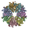 1pznS C: citing same article ( S: Starting model for refinement |
|---|---|
| Similar structure data |


-Links
 PDBj
PDBj


- Assembly
| Deposited unit | 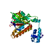
| ||||||||
|---|---|---|---|---|---|---|---|---|---|
| 1 |
| ||||||||
| Unit cell |
|
-Components
| #1: Protein | Mass: 35198.930 Da / Num. of mol.: 1 / Mutation: S2G Source method: isolated from a genetically manipulated source Source: (gene. exp.) Methanococcus voltae (archaea) / Gene: RADA / Plasmid: PET28B / Production host:  | ||||||
|---|---|---|---|---|---|---|---|
| #2: Chemical |   Mass: 24.305 Da / Num. of mol.: 2 / Source method: obtained synthetically / Formula: Mg Mass: 24.305 Da / Num. of mol.: 2 / Source method: obtained synthetically / Formula: Mg#3: Chemical |   Mass: 39.098 Da / Num. of mol.: 3 / Source method: obtained synthetically / Formula: K Mass: 39.098 Da / Num. of mol.: 3 / Source method: obtained synthetically / Formula: K#4: Chemical | ChemComp-ANP / |   Mass: 506.196 Da / Num. of mol.: 1 / Source method: obtained synthetically / Formula: C10H17N6O12P3 / Comment: AMP-PNP, energy-carrying molecule analogue*YM Mass: 506.196 Da / Num. of mol.: 1 / Source method: obtained synthetically / Formula: C10H17N6O12P3 / Comment: AMP-PNP, energy-carrying molecule analogue*YM#5: Water | ChemComp-HOH / |  Mass: 18.015 Da / Num. of mol.: 100 / Source method: isolated from a natural source / Formula: H2O Mass: 18.015 Da / Num. of mol.: 100 / Source method: isolated from a natural source / Formula: H2O |
-Experimental details
-Experiment
| Experiment | Method: X-RAY DIFFRACTION / Number of used crystals: 1 |
|---|
- Sample preparation
| Crystal | Density Matthews: 2.98 Å3/Da / Density % sol: 58.4 % |
|---|---|
| Crystal grow | Temperature: 294 K / Method: vapor diffusion, hanging drop / pH: 7.4 Details: 7% PEG 3350, 50 MM HEPES BUFFER, 50 MM MGCL2, 0.6 M K AC, pH 7.40, VAPOR DIFFUSION, HANGING DROP, temperature 294K |
-Data collection
| Diffraction | Mean temperature: 100 K |
|---|---|
| Diffraction source | Source: ROTATING ANODE / Type: ENRAF-NONIUS / Wavelength: 1.5418 / Wavelength: 1.5418 Å |
| Detector | Type: ENRAF-NONIUS / Detector: CCD / Date: Jan 16, 2004 / Details: MIRRORS |
| Radiation | Monochromator: BRUKERS MIRRORS / Protocol: SINGLE WAVELENGTH / Monochromatic (M) / Laue (L): M / Scattering type: x-ray |
| Radiation wavelength | Wavelength: 1.5418 Å / Relative weight: 1 |
| Reflection | Resolution: 2→40 Å / Num. all: 28274 / Num. obs: 28217 / % possible obs: 99.8 % / Observed criterion σ(F): 0 / Observed criterion σ(I): 0 / Redundancy: 5.5 % / Rmerge(I) obs: 0.039 / Rsym value: 0.039 / Net I/σ(I): 14.9 |
| Reflection shell | Resolution: 2→2.1 Å / Rmerge(I) obs: 0.279 / Rsym value: 0.279 / % possible all: 99.1 |
- Processing
| Software |
| ||||||||||||||||||||||||||||||||||||||||||||||||||||||||||||
|---|---|---|---|---|---|---|---|---|---|---|---|---|---|---|---|---|---|---|---|---|---|---|---|---|---|---|---|---|---|---|---|---|---|---|---|---|---|---|---|---|---|---|---|---|---|---|---|---|---|---|---|---|---|---|---|---|---|---|---|---|---|
| Refinement | Method to determine structure: MOLECULAR REPLACEMENT Starting model: PDB ENTRY 1PZN Resolution: 2→20 Å / Isotropic thermal model: ISOTROPIC / Cross valid method: THROUGHOUT / σ(F): 1 / Stereochemistry target values: Engh & Huber / Details: CNS REFINEMENT
| ||||||||||||||||||||||||||||||||||||||||||||||||||||||||||||
| Refinement step | Cycle: LAST / Resolution: 2→20 Å
| ||||||||||||||||||||||||||||||||||||||||||||||||||||||||||||
| Refine LS restraints |
| ||||||||||||||||||||||||||||||||||||||||||||||||||||||||||||
| Xplor file |
|
