 Movie
Movie Controller

[English] 日本語
 Yorodumi
- PDB-2fmf: Crystal structure of CheY in complex with CheZ 200-214 solved fro... -
+ Open data

- Basic information
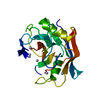
| Entry | Database: PDB / ID: 2fmf | ||||||
|---|---|---|---|---|---|---|---|
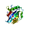
| Title | Crystal structure of CheY in complex with CheZ 200-214 solved from a F432 crystal grown in Hepes (pH 7.5) | ||||||
 Components Components |
| ||||||
 Keywords Keywords | SIGNALING PROTEIN / CHEMOTAXIS / BEF(3)(-)-BOUND CHEY / CHEY-CHEZ PEPTIDE COMPLEX | ||||||
| Function / homology |  Function and homology information Function and homology informationarchaeal or bacterial-type flagellum-dependent cell motility / regulation of chemotaxis / Hydrolases; Acting on ester bonds; Phosphoric-monoester hydrolases / bacterial-type flagellum / phosphorelay signal transduction system / phosphoprotein phosphatase activity / chemotaxis / metal ion binding / cytoplasm Similarity search - Function | ||||||
| Biological species |  Salmonella typhimurium (bacteria) Salmonella typhimurium (bacteria) | ||||||
| Method |  X-RAY DIFFRACTION / SYNCHROTRON / MOLECULAR REPLACEMENT / Resolution: 1.998 Å X-RAY DIFFRACTION / SYNCHROTRON / MOLECULAR REPLACEMENT / Resolution: 1.998 Å | ||||||
 Authors Authors | Guhaniyogi, J. / Robinson, V.L. / Stock, A.M. | ||||||
 Citation Citation | Journal: J.Mol.Biol. / Year: 2006 Title: Crystal Structures of Beryllium Fluoride-free and Beryllium Fluoride-bound CheY in Complex with the Conserved C-terminal Peptide of CheZ Reveal Dual Binding Modes Specific to CheY Conformation. Authors: Guhaniyogi, J. / Robinson, V.L. / Stock, A.M. | ||||||
| History |
|
- Structure visualization
| Structure viewer | Molecule: MolmilJmol/JSmol |
|---|
- Downloads & links
-Download
| PDBx/mmCIF format | 2fmf.cif.gz | 42 KB | Display | PDBx/mmCIF format |
|---|---|---|---|---|
| PDB format | pdb2fmf.ent.gz | 31.2 KB | Display | PDB format |
| PDBx/mmJSON format | 2fmf.json.gz | Tree view | PDBx/mmJSON format | |
| Others |  Other downloads Other downloads |
-Validation report
| Arichive directory | https://data.pdbj.org/pub/pdb/validation_reports/fm/2fmfftp://data.pdbj.org/pub/pdb/validation_reports/fm/2fmf | HTTPS FTP |
|---|
-Related structure data
| Related structure data |  2fkaC  2flkC  2flwC  2fmhC  2fmiC  2fmkC  1fqwS C: citing same article ( S: Starting model for refinement |
|---|---|
| Similar structure data |


-Links
 PDBj
PDBj

- Assembly
| Deposited unit | 
| ||||||||||||
|---|---|---|---|---|---|---|---|---|---|---|---|---|---|
| 1 |
| ||||||||||||
| Unit cell |
| ||||||||||||
| Components on special symmetry positions |
|
-Components
| #1: Protein | Mass: 14140.385 Da / Num. of mol.: 1 Source method: isolated from a genetically manipulated source Source: (gene. exp.) Salmonella typhimurium (bacteria) / Strain: LT2 / Gene: cheY / Plasmid: pUC18 / Production host: |
|---|---|
| #2: Protein/peptide | Mass: 1622.687 Da / Num. of mol.: 1 / Fragment: residues 200-214 / Source method: obtained synthetically Details: This sequence corresponds to the C-terminal 15 residues of the CheZ protein occurring naturally in Salmonella enterica serovar Typhumurium References: UniProt: P07800 |
| #3: Chemical | ChemComp-SO4 /   Mass: 96.063 Da / Num. of mol.: 1 / Source method: obtained synthetically / Formula: SO4 Mass: 96.063 Da / Num. of mol.: 1 / Source method: obtained synthetically / Formula: SO4 |
| #4: Water | ChemComp-HOH /  Mass: 18.015 Da / Num. of mol.: 86 / Source method: isolated from a natural source / Formula: H2O Mass: 18.015 Da / Num. of mol.: 86 / Source method: isolated from a natural source / Formula: H2O |
-Experimental details
-Experiment
| Experiment | Method: X-RAY DIFFRACTION / Number of used crystals: 1 |
|---|
- Sample preparation
| Crystal grow | Temperature: 298 K / Method: vapor diffusion, hanging drop / pH: 7.5 Details: 2M ammonium sulfate, 0.2M lithium sulfate, 0.1M Hepes, pH 7.5, VAPOR DIFFUSION, HANGING DROP, temperature 298.0K |
|---|
-Data collection
| Diffraction | Mean temperature: 100 K |
|---|---|
| Diffraction source | Source: SYNCHROTRON / Site: NSLS  / Beamline: X4A / Wavelength: 0.9793 Å / Beamline: X4A / Wavelength: 0.9793 Å |
| Detector | Type: ADSC QUANTUM 4 / Detector: CCD / Date: Feb 17, 2003 |
| Radiation | Monochromator: KOHZU double crystal monochromator with a sagittally focused second crystal. Crystal type Si(111) Protocol: SINGLE WAVELENGTH / Monochromatic (M) / Laue (L): M / Scattering type: x-ray |
| Radiation wavelength | Wavelength: 0.9793 Å / Relative weight: 1 |
| Reflection | Resolution: 1.998→20 Å / Num. all: 22840 / Num. obs: 22136 / % possible obs: 97.1 % / Observed criterion σ(I): -3 / Biso Wilson estimate: 34.1 Å2 / Rsym value: 0.078 / Net I/σ(I): 22 |
| Reflection shell | Resolution: 1.998→2.07 Å / Mean I/σ(I) obs: 6.6 / Num. unique all: 2148 / Rsym value: 0.251 / % possible all: 97 |
- Processing
| Software |
| |||||||||||||||||||||||||||||||||||||||||||||||||||||||||||||||||||||||||||||||||||||||||||||||||||||||||
|---|---|---|---|---|---|---|---|---|---|---|---|---|---|---|---|---|---|---|---|---|---|---|---|---|---|---|---|---|---|---|---|---|---|---|---|---|---|---|---|---|---|---|---|---|---|---|---|---|---|---|---|---|---|---|---|---|---|---|---|---|---|---|---|---|---|---|---|---|---|---|---|---|---|---|---|---|---|---|---|---|---|---|---|---|---|---|---|---|---|---|---|---|---|---|---|---|---|---|---|---|---|---|---|---|---|---|
| Refinement | Method to determine structure: MOLECULAR REPLACEMENT Starting model: PDB ENTRY 1FQW Resolution: 1.998→19.8 Å / Cor.coef. Fo:Fc: 0.946 / Cor.coef. Fo:Fc free: 0.942 / SU B: 3.483 / SU ML: 0.1 / Cross valid method: THROUGHOUT / σ(F): 0 / ESU R: 0.12 / ESU R Free: 0.111 / Stereochemistry target values: MAXIMUM LIKELIHOOD / Details: HYDROGENS HAVE BEEN ADDED IN THE RIDING POSITIONS
| |||||||||||||||||||||||||||||||||||||||||||||||||||||||||||||||||||||||||||||||||||||||||||||||||||||||||
| Solvent computation | Ion probe radii: 0.8 Å / Shrinkage radii: 0.8 Å / VDW probe radii: 1.4 Å / Solvent model: BABINET MODEL WITH MASK | |||||||||||||||||||||||||||||||||||||||||||||||||||||||||||||||||||||||||||||||||||||||||||||||||||||||||
| Displacement parameters | Biso mean: 27.445 Å2 | |||||||||||||||||||||||||||||||||||||||||||||||||||||||||||||||||||||||||||||||||||||||||||||||||||||||||
| Refinement step | Cycle: LAST / Resolution: 1.998→19.8 Å
| |||||||||||||||||||||||||||||||||||||||||||||||||||||||||||||||||||||||||||||||||||||||||||||||||||||||||
| Refine LS restraints |
| |||||||||||||||||||||||||||||||||||||||||||||||||||||||||||||||||||||||||||||||||||||||||||||||||||||||||
| LS refinement shell | Resolution: 1.998→2.05 Å / Total num. of bins used: 20
|
