 Movie
Movie Controller

[English] 日本語
 Yorodumi
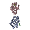
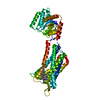
- PDB-2fgs: Crystal structure of Campylobacter jejuni YCEI protein, structura... -
+ Open data

- Basic information
| Entry | Database: PDB / ID: 2fgs | ||||||
|---|---|---|---|---|---|---|---|
| Title | Crystal structure of Campylobacter jejuni YCEI protein, structural genomics | ||||||
 Components Components | Putative periplasmic protein | ||||||
 Keywords Keywords | LIPID BINDING PROTEIN / STRUCTURAL GENOMICS / PROTEIN STRUCTURE INITIATIVE / PSI / NYSGXRC / New York SGX Research Center for Structural Genomics | ||||||
| Function / homology |  Function and homology information Function and homology informationLipid/polyisoprenoid-binding, YceI-like / Lipid/polyisoprenoid-binding, YceI-like / Lipid/polyisoprenoid-binding, YceI-like superfamily / YceI-like domain / YceI-like domain / Lipocalin / Beta Barrel / Mainly Beta Similarity search - Domain/homology | ||||||
| Biological species |   Campylobacter jejuni (Campylobacter) Campylobacter jejuni (Campylobacter) | ||||||
| Method |  X-RAY DIFFRACTION / MOLECULAR REPLACEMENT / Resolution: 2.9 Å X-RAY DIFFRACTION / MOLECULAR REPLACEMENT / Resolution: 2.9 Å | ||||||
 Authors Authors | Patskovsky, Y. / Ramagopal, U. / Almo, S.C. / Burley, S.K. / New York SGX Research Center for Structural Genomics (NYSGXRC) | ||||||
 Citation Citation | Journal: To be Published Title: Crystal Structure of Campylobacter Jejuni YceI Periplasmic Protein Authors: Patskovsky, Y. / Ramagopal, U. / Almo, S.C. | ||||||
| History |
|
- Structure visualization
| Structure viewer | Molecule: MolmilJmol/JSmol |
|---|
- Downloads & links
-Download
| PDBx/mmCIF format | 2fgs.cif.gz | 46.1 KB | Display | PDBx/mmCIF format |
|---|---|---|---|---|
| PDB format | pdb2fgs.ent.gz | 32.1 KB | Display | PDB format |
| PDBx/mmJSON format | 2fgs.json.gz | Tree view | PDBx/mmJSON format | |
| Others |  Other downloads Other downloads |
-Validation report
| Arichive directory | https://data.pdbj.org/pub/pdb/validation_reports/fg/2fgsftp://data.pdbj.org/pub/pdb/validation_reports/fg/2fgs | HTTPS FTP |
|---|
-Related structure data
| Related structure data |  1y0gS S: Starting model for refinement |
|---|---|
| Similar structure data | |
| Other databases |


-Links
 PDBj
PDBj- Assembly
| Deposited unit | 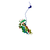
| ||||||||
|---|---|---|---|---|---|---|---|---|---|
| 1 | 
| ||||||||
| Unit cell |
| ||||||||
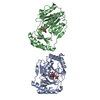
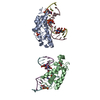
| Details | The biological assembly is a homodimer. The second part of the assembly is generated by the two-fold axis -x, y-x, 1-z |
-Components
| #1: Protein | Mass: 20068.596 Da / Num. of mol.: 1 Source method: isolated from a genetically manipulated source Source: (gene. exp.) Campylobacter jejuni (Campylobacter) / Gene: Cj0420 / Plasmid: pET / Species (production host): Escherichia coli / Production host: |
|---|---|
| #2: Chemical | ChemComp-SO4 /   Mass: 96.063 Da / Num. of mol.: 1 / Source method: obtained synthetically / Formula: SO4 Mass: 96.063 Da / Num. of mol.: 1 / Source method: obtained synthetically / Formula: SO4 |
-Experimental details
-Experiment
| Experiment | Method: X-RAY DIFFRACTION / Number of used crystals: 1 |
|---|
- Sample preparation
| Crystal | Density Matthews: 6.1 Å3/Da / Density % sol: 78.25 % |
|---|---|
| Crystal grow | Temperature: 290 K / Method: vapor diffusion, sitting drop / pH: 6.5 Details: 3.2M AMMONIUM SULFATE, 0.1M BIS-TRIS, pH 6.50, VAPOR DIFFUSION, SITTING DROP, temperature 290K |
-Data collection
| Diffraction | Mean temperature: 100 K |
|---|---|
| Diffraction source | Source: ROTATING ANODE / Type: RIGAKU RU200 / Wavelength: 1.5418 Å |
| Detector | Type: RIGAKU RAXIS IV / Detector: IMAGE PLATE / Date: Dec 19, 2005 / Details: MIRRORS |
| Radiation | Monochromator: MIRRORS / Protocol: SINGLE WAVELENGTH / Monochromatic (M) / Laue (L): M / Scattering type: x-ray |
| Radiation wavelength | Wavelength: 1.5418 Å / Relative weight: 1 |
| Reflection | Resolution: 2.9→50 Å / Num. all: 11129 / Num. obs: 11129 / % possible obs: 99.8 % / Observed criterion σ(F): 0 / Observed criterion σ(I): 0 / Redundancy: 14.1 % / Rmerge(I) obs: 0.124 / Rsym value: 0.088 / Net I/σ(I): 6.2 |
| Reflection shell | Resolution: 2.9→3 Å / Redundancy: 13.8 % / Rmerge(I) obs: 0.398 / Mean I/σ(I) obs: 2.2 / Num. unique all: 1092 / Rsym value: 0.46 / % possible all: 100 |
- Processing
| Software |
| ||||||||||||||||||||||||||||||||||||||||||||||||||||||||||||||||||||||||||||||||||||||||||
|---|---|---|---|---|---|---|---|---|---|---|---|---|---|---|---|---|---|---|---|---|---|---|---|---|---|---|---|---|---|---|---|---|---|---|---|---|---|---|---|---|---|---|---|---|---|---|---|---|---|---|---|---|---|---|---|---|---|---|---|---|---|---|---|---|---|---|---|---|---|---|---|---|---|---|---|---|---|---|---|---|---|---|---|---|---|---|---|---|---|---|---|
| Refinement | Method to determine structure: MOLECULAR REPLACEMENT Starting model: PDB ENTRY 1Y0G Resolution: 2.9→20 Å / Cor.coef. Fo:Fc: 0.92 / Cor.coef. Fo:Fc free: 0.913 / SU B: 12.635 / SU ML: 0.238 / Cross valid method: THROUGHOUT / σ(F): 0 / ESU R: 0.364 / ESU R Free: 0.294 / Stereochemistry target values: MAXIMUM LIKELIHOOD Details: HYDROGENS HAVE BEEN ADDED IN THE RIDING POSITIONS UNIDENTIFIED DENSITY WAS FOUND IN THE CENTRAL CAVITY OF THE PROTEIN BUT THE STRUCTURE OF THE BOUND LIGAND HAS NOT BEEN DETERMINED YET
| ||||||||||||||||||||||||||||||||||||||||||||||||||||||||||||||||||||||||||||||||||||||||||
| Solvent computation | Ion probe radii: 0.8 Å / Shrinkage radii: 0.8 Å / VDW probe radii: 1.2 Å / Solvent model: MASK | ||||||||||||||||||||||||||||||||||||||||||||||||||||||||||||||||||||||||||||||||||||||||||
| Displacement parameters | Biso mean: 69.73 Å2
| ||||||||||||||||||||||||||||||||||||||||||||||||||||||||||||||||||||||||||||||||||||||||||
| Refinement step | Cycle: LAST / Resolution: 2.9→20 Å
| ||||||||||||||||||||||||||||||||||||||||||||||||||||||||||||||||||||||||||||||||||||||||||
| Refine LS restraints |
| ||||||||||||||||||||||||||||||||||||||||||||||||||||||||||||||||||||||||||||||||||||||||||
| LS refinement shell | Resolution: 2.9→2.974 Å / Rfactor Rfree error: 0.043 / Total num. of bins used: 20
|
