 Movie
Movie Controller

[English] 日本語
 Yorodumi
- PDB-2fb5: Structural Genomics; The crystal structure of the hypothetical me... -
+ Open data

- Basic information
| Entry | Database: PDB / ID: 2fb5 | ||||||
|---|---|---|---|---|---|---|---|
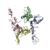
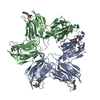
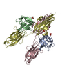
| Title | Structural Genomics; The crystal structure of the hypothetical membrane spanning protein from Bacillus cereus | ||||||
 Components Components | hypothetical Membrane Spanning Protein | ||||||
 Keywords Keywords | MEMBRANE PROTEIN / Structural Genomics / PSI / Protein Structure Initiative / Midwest Center for Structural Genomics / MCSG | ||||||
| Function / homology |  Function and homology information Function and homology informationdiadenylate cyclase / diadenylate cyclase activity / adenylate cyclase activity / cAMP biosynthetic process / ATP binding Similarity search - Function | ||||||
| Biological species |  | ||||||
| Method |  X-RAY DIFFRACTION / SYNCHROTRON / SAD / Resolution: 1.99 Å X-RAY DIFFRACTION / SYNCHROTRON / SAD / Resolution: 1.99 Å | ||||||
 Authors Authors | Zhang, R. / Zhou, M. / Ginell, S. / Abdullah, J. / Collart, F. / Joachimiak, A. / Midwest Center for Structural Genomics (MCSG) | ||||||
 Citation Citation | Journal: To be Published Title: The crystal structure of the hypothetical membrane spanning protein from Bacillus cereus Authors: Zhang, R. / Zhou, M. / Ginell, S. / Abdullah, J. / Collart, F. / Joachimiak, A. | ||||||
| History |
|
- Structure visualization
| Structure viewer | Molecule: MolmilJmol/JSmol |
|---|
- Downloads & links
-Download
| PDBx/mmCIF format | 2fb5.cif.gz | 144.8 KB | Display | PDBx/mmCIF format |
|---|---|---|---|---|
| PDB format | pdb2fb5.ent.gz | 115.3 KB | Display | PDB format |
| PDBx/mmJSON format | 2fb5.json.gz | Tree view | PDBx/mmJSON format | |
| Others |  Other downloads Other downloads |
-Validation report
| Arichive directory | https://data.pdbj.org/pub/pdb/validation_reports/fb/2fb5ftp://data.pdbj.org/pub/pdb/validation_reports/fb/2fb5 | HTTPS FTP |
|---|
-Related structure data
| Similar structure data | |
|---|---|
| Other databases |


-Links
 PDBj
PDBj- Assembly
| Deposited unit | 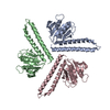
| ||||||||||||
|---|---|---|---|---|---|---|---|---|---|---|---|---|---|
| 1 |
| ||||||||||||
| 2 | 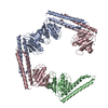
| ||||||||||||
| 3 | 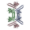
| ||||||||||||
| Unit cell |
| ||||||||||||
| Components on special symmetry positions |
| ||||||||||||
| Details | This protein existed as trimer. The molecules A, B, C represent the trimer in the asymmetric unit. |
-Components
| #1: Protein | Mass: 22574.748 Da / Num. of mol.: 3 Source method: isolated from a genetically manipulated source Source: (gene. exp.) #2: Water | ChemComp-HOH / |  Mass: 18.015 Da / Num. of mol.: 846 / Source method: isolated from a natural source / Formula: H2O Mass: 18.015 Da / Num. of mol.: 846 / Source method: isolated from a natural source / Formula: H2O |
|---|
-Experimental details
-Experiment
| Experiment | Method: X-RAY DIFFRACTION / Number of used crystals: 1 |
|---|
- Sample preparation
| Crystal | Density Matthews: 3.44 Å3/Da / Density % sol: 64.2 % |
|---|---|
| Crystal grow | Temperature: 287 K / Method: vapor diffusion, hanging drop / pH: 7 Details: 0.18M Sodium Malonate, 18% PEG3350, pH 7.0, VAPOR DIFFUSION, HANGING DROP, temperature 287K |
-Data collection
| Diffraction | Mean temperature: 100 K |
|---|---|
| Diffraction source | Source: SYNCHROTRON / Site: APS  / Beamline: 19-ID / Wavelength: 0.9798 Å / Beamline: 19-ID / Wavelength: 0.9798 Å |
| Detector | Type: ADSC QUANTUM 4 / Detector: CCD / Date: Nov 14, 2004 / Details: mirrors |
| Radiation | Monochromator: Si 111 channel / Protocol: SINGLE WAVELENGTH / Monochromatic (M) / Laue (L): M / Scattering type: x-ray |
| Radiation wavelength | Wavelength: 0.9798 Å / Relative weight: 1 |
| Reflection | Resolution: 1.99→77.62 Å / Num. all: 62291 / Num. obs: 61484 / % possible obs: 97.2 % / Observed criterion σ(F): 2 / Observed criterion σ(I): 2 / Redundancy: 8.1 % / Biso Wilson estimate: 30.92 Å2 / Rmerge(I) obs: 0.09 / Net I/σ(I): 26 |
| Reflection shell | Resolution: 2→2.07 Å / Redundancy: 4.9 % / Rmerge(I) obs: 0.489 / Mean I/σ(I) obs: 1.51 / Num. unique all: 7786 / % possible all: 73.1 |
- Processing
| Software |
| ||||||||||||||||||||||||||||||||||||||||||||||||||||||||||||||||||||||||||||||||||||||||||
|---|---|---|---|---|---|---|---|---|---|---|---|---|---|---|---|---|---|---|---|---|---|---|---|---|---|---|---|---|---|---|---|---|---|---|---|---|---|---|---|---|---|---|---|---|---|---|---|---|---|---|---|---|---|---|---|---|---|---|---|---|---|---|---|---|---|---|---|---|---|---|---|---|---|---|---|---|---|---|---|---|---|---|---|---|---|---|---|---|---|---|---|
| Refinement | Method to determine structure: SAD / Resolution: 1.99→77.62 Å / Cor.coef. Fo:Fc: 0.955 / Cor.coef. Fo:Fc free: 0.937 / SU B: 5.966 / SU ML: 0.09 / TLS residual ADP flag: LIKELY RESIDUAL / Cross valid method: THROUGHOUT / σ(F): 0 / σ(I): 0 / ESU R: 0.145 / ESU R Free: 0.136 Stereochemistry target values: MAXIMUM LIKELIHOOD WITH PHASES Details: HYDROGENS HAVE BEEN ADDED IN THE RIDING POSITIONS
| ||||||||||||||||||||||||||||||||||||||||||||||||||||||||||||||||||||||||||||||||||||||||||
| Solvent computation | Ion probe radii: 0.8 Å / Shrinkage radii: 0.8 Å / VDW probe radii: 1.2 Å / Solvent model: MASK | ||||||||||||||||||||||||||||||||||||||||||||||||||||||||||||||||||||||||||||||||||||||||||
| Displacement parameters | Biso mean: 30.915 Å2
| ||||||||||||||||||||||||||||||||||||||||||||||||||||||||||||||||||||||||||||||||||||||||||
| Refine analyze |
| ||||||||||||||||||||||||||||||||||||||||||||||||||||||||||||||||||||||||||||||||||||||||||
| Refinement step | Cycle: LAST / Resolution: 1.99→77.62 Å
| ||||||||||||||||||||||||||||||||||||||||||||||||||||||||||||||||||||||||||||||||||||||||||
| Refine LS restraints |
| ||||||||||||||||||||||||||||||||||||||||||||||||||||||||||||||||||||||||||||||||||||||||||
| LS refinement shell | Resolution: 1.994→2.046 Å / Total num. of bins used: 20
| ||||||||||||||||||||||||||||||||||||||||||||||||||||||||||||||||||||||||||||||||||||||||||
| Refinement TLS params. | Method: refined / Origin x: 39.419 Å / Origin y: 103.765 Å / Origin z: 27.804 Å
| ||||||||||||||||||||||||||||||||||||||||||||||||||||||||||||||||||||||||||||||||||||||||||
| Refinement TLS group |
|
