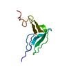
- PDB-2eze: SOLUTION STRUCTURE OF A COMPLEX OF THE SECOND DNA BINDING DOMAIN ... -
+
Open data
ID or keywords:
Loading...
-
Basic information
Entry
Database: PDB / ID: 2eze
Title
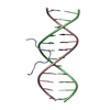
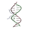
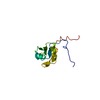
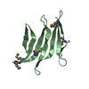
SOLUTION STRUCTURE OF A COMPLEX OF THE SECOND DNA BINDING DOMAIN OF HUMAN HMG-I(Y) BOUND TO DNA DODECAMER CONTAINING THE PRDII SITE OF THE INTERFERON-BETA PROMOTER, NMR, 35 STRUCTURES
Components
DNA (5'-D(*GP*AP*GP*GP*AP*AP*TP*TP*TP*CP*CP*C)-3')
DNA (5'-D(*GP*GP*GP*AP*AP*AP*TP*TP*CP*CP*TP*C)-3')
HIGH MOBILITY GROUP PROTEIN HMG-I/HMG-Y
Keywords
DNA BINDING PROTEIN/DNA / DNA BINDING PROTEIN / MINOR GROOVE DNA BINDING / TRANSCRIPTIONAL CO-ACTIVATOR / ARCHITECTURAL FACTOR / COMPLEX (DNA-BINDING PROTEIN-DNA) / DNA BINDING PROTEIN-DNA COMPLEX
Function / homology
Function and homology information
senescence-associated heterochromatin focus / nuclear retinoic acid receptor binding / oncogene-induced cell senescence / peroxisome proliferator activated receptor binding / nucleosome disassembly / Integration of viral DNA into host genomic DNA / Autointegration results in viral DNA circles / DNA binding, bending / Formation of Senescence-Associated Heterochromatin Foci (SAHF) / 2-LTR circle formation ...senescence-associated heterochromatin focus / nuclear retinoic acid receptor binding / oncogene-induced cell senescence / peroxisome proliferator activated receptor binding / nucleosome disassembly / Integration of viral DNA into host genomic DNA / Autointegration results in viral DNA circles / DNA binding, bending / Formation of Senescence-Associated Heterochromatin Foci (SAHF) / 2-LTR circle formation / Vpr-mediated nuclear import of PICs / Integration of provirus / APOBEC3G mediated resistance to HIV-1 infection / minor groove of adenine-thymine-rich DNA binding / 5'-deoxyribose-5-phosphate lyase activity / nuclear retinoid X receptor binding / cis-regulatory region sequence-specific DNA binding / DNA-(apurinic or apyrimidinic site) endonuclease activity / molecular function activator activity / transcription coregulator binding / transcription coregulator activity / base-excision repair / RNA polymerase II transcription regulator complex / structural constituent of chromatin / transcription regulator complex / nuclear membrane / molecular adaptor activity / transcription coactivator activity / intracellular signal transduction / RNA polymerase II cis-regulatory region sequence-specific DNA binding / negative regulation of cell population proliferation / focal adhesion / negative regulation of DNA-templated transcription / chromatin binding / regulation of DNA-templated transcription / positive regulation of DNA-templated transcription / enzyme binding / positive regulation of transcription by RNA polymerase II / DNA binding / RNA binding / nucleoplasm / nucleus / cytosol Similarity search - Function
High mobility group protein HMGA / HMG-I/HMG-Y, DNA-binding, conserved site / HMG-I and HMG-Y DNA-binding domain (A+T-hook). / DNA binding domain with preference for A/T rich regions / AT hook, DNA-binding motif Similarity search - Domain/homology
DNA (5'-D(*GP*GP*GP*AP*AP*AP*TP*TP*CP*CP*TP*C)-3')
Mass: 3662.404 Da / Num. of mol.: 1 / Source method: obtained synthetically
#2: DNA chain
DNA (5'-D(*GP*AP*GP*GP*AP*AP*TP*TP*TP*CP*CP*C)-3')
Mass: 3662.404 Da / Num. of mol.: 1 / Source method: obtained synthetically
#3: Protein/peptide
HIGHMOBILITYGROUPPROTEINHMG-I/HMG-Y / HIGH MOBILITY GROUP PROTEIN HMG
Mass: 2727.262 Da / Num. of mol.: 1 Source method: isolated from a genetically manipulated source Source: (gene. exp.) Homo sapiens (human) / Gene: POTENTIAL / Production host: Escherichia coli (E. coli) / References: UniProt: P17096
-
Experimental details
-
Experiment
Experiment
Method: SOLUTION NMR
NMR details
Text: DATA WERE RECORDED ON A 2:1 COMPLEX OF DNA DODECAMER TO HMG-I(Y) 50-91 WHICH CONTAINS THE SECOND AND THIRD DNA DNA BINDING DOMAINS. EACH DNA BINDING DOMAIN BINDS TO 1 MOLECULE OF DNA.
-
Sample preparation
Sample conditions
pH: 6.1 / Temperature: 306 K
Crystal grow
*PLUS
Method: other / Details: NMR
-
NMR measurement
NMR spectrometer
Type
Manufacturer
Model
Field strength (MHz)
Spectrometer-ID
Bruker AMX500
Bruker
AMX500
600
1
Bruker AMX600
Bruker
AMX600
500
2
Bruker DMX600
Bruker
DMX600
750
3
Bruker DMX750
Bruker
DMX750
750
4
-
Processing
Software
Name
Version
Classification
X-PLOR
3.1
modelbuilding
X-PLOR
3.1
refinement
X-PLOR
3.1
phasing
NMR software
Name
Version
Developer
Classification
X-PLOR
3.1
BRUNGER
refinement
XPLOR MODIFIED
MODIFIED
structuresolution
Refinement
Method: simulated annealing / Software ordinal: 1 Details: THE STRUCTURES WERE CALCULATED USING THE SIMULATED ANNEALING PROTOCOL OF NILGES ET AL. (1988) FEBS LETT. 229, 129 - 136 USING THE PROGRAM X-PLOR 3.1 (BRUNGER) MODIFIED TO INCORPORATE ...Details: THE STRUCTURES WERE CALCULATED USING THE SIMULATED ANNEALING PROTOCOL OF NILGES ET AL. (1988) FEBS LETT. 229, 129 - 136 USING THE PROGRAM X-PLOR 3.1 (BRUNGER) MODIFIED TO INCORPORATE COUPLING CONSTANT (GARRETT ET AL. (1984) J. MAGN RESON. SERIES B 104, 99 - 103), CARBON CHEMICAL SHIFT (KUSZEWSKI ET AL. (1995) J. MAGN. RESON. SERIES B 106, 92 - 96) RESTRAINTS AND A CONFORMATIONAL DATABASE POTENTIAL (KUSZEWSKI ET AL. (1996) PROTEIN SCI 5, 1067 - 1080 AND (1997) J. MAGN. RESON. 125, 171-177) THE 3D STRUCTURE OF THE COMPLEX OF THE SECOND DNA BINDING DOMAIN OF HMG-I(Y) COMPLEXED TO DNA WAS SOLVED BY MULTI-DIMENSIONAL HETERONUCLEAR-EDITED AND -FILTERED NMR (A) PROTEIN: 71 SEQUENTIAL (|I-J|=1), 4 MEDIUM RANGE (1 < |I-J| >=5) AND 64 INTRARESIDUE APPROXIMATE INTERPROTON DISTANCE RESTRAINTS; NULL 36 TORSION ANGLE RESTRAINTS 13 THREE-BOND HN-HA AND 8 THREE_BOND COCO COUPLING CONSTANT RESTRAINTS; 39 (21 CALPHA AND 18 CBETA) 13C SHIFT RESTRAINTS. (B) DNA: 249 INTRARESIDUE, 119 SEQUENTIAL INTRASTRAND AND 33 INTERSTRAND INTERPROTON DISTANCE RESTRAINTS; 42 DISTANCES FOR WATSON-CRICK BASE PAIR HYDROGEN BONDS; 136 TORSION ANGLE RESTRAINTS (C) 73 INTERMOLECULAR INTERPROTON DISTANCE RESTRAINTS (D) 5 INTERMOLECULAR DISTANCE RESTRAINTS TO PHOSPHATES (E) 20 'REPULSIVE' RESTRAINTS THE STRUCTURE IN THIS ENTRY ARE THE 35 INDIVIDUAL SIMULATED ANNEALING STRUCTURES. THE RESTRAINED REGULARIZED MEAN STRUCTURE IS FOUND IN PDB ENTRY 2EZD. THE LAST NUMERIC COLUMN IN THE INDIVIDUAL SA STRUCTURES HAS NO MEANING. RESIDUES 3 - 27 OF THE PROTEIN CORRESPOND TO RESIDUES 51 - 75 OF INTACT HMG-I(Y). RESIDUES 3 - 5 AND 20 - 27 ARE DISORDERED.
NMR ensemble
Conformers calculated total number: 35 / Conformers submitted total number: 35
+
About Yorodumi
-
News
-
Feb 9, 2022. New format data for meta-information of EMDB entries
New format data for meta-information of EMDB entries
Version 3 of the EMDB header file is now the official format.
The previous official version 1.9 will be removed from the archive.
In the structure databanks used in Yorodumi, some data are registered as the other names, "COVID-19 virus" and "2019-nCoV". Here are the details of the virus and the list of structure data.
Jan 31, 2019. EMDB accession codes are about to change! (news from PDBe EMDB page)
EMDB accession codes are about to change! (news from PDBe EMDB page)
The allocation of 4 digits for EMDB accession codes will soon come to an end. Whilst these codes will remain in use, new EMDB accession codes will include an additional digit and will expand incrementally as the available range of codes is exhausted. The current 4-digit format prefixed with “EMD-” (i.e. EMD-XXXX) will advance to a 5-digit format (i.e. EMD-XXXXX), and so on. It is currently estimated that the 4-digit codes will be depleted around Spring 2019, at which point the 5-digit format will come into force.
The EM Navigator/Yorodumi systems omit the EMD- prefix.
Related info.:Q: What is EMD? / ID/Accession-code notation in Yorodumi/EM Navigator
Yorodumi is a browser for structure data from EMDB, PDB, SASBDB, etc.
This page is also the successor to EM Navigator detail page, and also detail information page/front-end page for Omokage search.
The word "yorodu" (or yorozu) is an old Japanese word meaning "ten thousand". "mi" (miru) is to see.
Related info.:EMDB / PDB / SASBDB / Comparison of 3 databanks / Yorodumi Search / Aug 31, 2016. New EM Navigator & Yorodumi / Yorodumi Papers / Jmol/JSmol / Function and homology information / Changes in new EM Navigator and Yorodumi
 Movie
Movie Controller

 Yorodumi
 Open data
 Basic information
 Components
 Keywords
 Function and homology information
 Homo sapiens (human)
 Authors
 Citation
 Structure visualization
 Downloads & links
 Other downloads
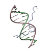


 PDBj
PDBj


 Assembly
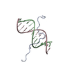

 Sample preparation
 Processing
 X-PLOR