 Movie
Movie Controller

[English] 日本語
 Yorodumi
- PDB-2ewe: Crystal structure of Pectate Lyase C R218K mutant in complex with... -
+ Open data

- Basic information
| Entry | Database: PDB / ID: 2ewe | |||||||||
|---|---|---|---|---|---|---|---|---|---|---|
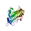
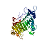
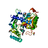
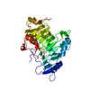
| Title | Crystal structure of Pectate Lyase C R218K mutant in complex with pentagalacturonic acid | |||||||||
 Components Components | Pectate lyase C | |||||||||
 Keywords Keywords | LYASE / parallel beta helix / protein-oligosaccharide interactions | |||||||||
| Function / homology |  Function and homology information Function and homology informationpectate lyase / pectate lyase activity / pectin catabolic process / extracellular region / metal ion binding Similarity search - Function | |||||||||
| Biological species |  Erwinia chrysanthemi (bacteria) Erwinia chrysanthemi (bacteria) | |||||||||
| Method |  X-RAY DIFFRACTION / SYNCHROTRON / FOURIER SYNTHESIS / Resolution: 2.2 Å X-RAY DIFFRACTION / SYNCHROTRON / FOURIER SYNTHESIS / Resolution: 2.2 Å | |||||||||
 Authors Authors | Scavetta, R.D. / Jurnak, F. | |||||||||
 Citation Citation | Journal: Plant Cell / Year: 1999 Title: Structure of a Plant Cell Wall Fragment Complexed to Pectate Lyase C Authors: Scavetta, R.D. / Herron, S.R. / Hotchkiss, A.T. / Kita, N. / Keen, N.T. / Benen, J.A. / Kester, H.C. / Visser, J. / Jurnak, F. | |||||||||
| History |
| |||||||||
| Remark 600 | HETEROGEN Only four of the five subunits of the pentagalacturonic acid were visible in the electron ...HETEROGEN Only four of the five subunits of the pentagalacturonic acid were visible in the electron density maps and could be modeled. |
- Structure visualization
| Structure viewer | Molecule: MolmilJmol/JSmol |
|---|
- Downloads & links
-Download
| PDBx/mmCIF format | 2ewe.cif.gz | 89.4 KB | Display | PDBx/mmCIF format |
|---|---|---|---|---|
| PDB format | pdb2ewe.ent.gz | 65.5 KB | Display | PDB format |
| PDBx/mmJSON format | 2ewe.json.gz | Tree view | PDBx/mmJSON format | |
| Others |  Other downloads Other downloads |
-Validation report
| Arichive directory | https://data.pdbj.org/pub/pdb/validation_reports/ew/2eweftp://data.pdbj.org/pub/pdb/validation_reports/ew/2ewe | HTTPS FTP |
|---|
-Related structure data
| Related structure data |  1airS S: Starting model for refinement |
|---|---|
| Similar structure data |


-Links
 PDBj
PDBj- Assembly
| Deposited unit | 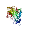
| ||||||||
|---|---|---|---|---|---|---|---|---|---|
| 1 |
| ||||||||
| Unit cell |
| ||||||||
| Details | The biological assembly is the monomer. |
-Components
| #1: Protein | Mass: 37705.590 Da / Num. of mol.: 1 / Mutation: R218K Source method: isolated from a genetically manipulated source Source: (gene. exp.) Erwinia chrysanthemi (bacteria) / Genus: Dickeya / Strain: EC16 / Gene: pelC / Plasmid: pRSET5A / Production host: | ||||
|---|---|---|---|---|---|
| #2: Polysaccharide | alpha-D-galactopyranuronic acid-(1-4)-alpha-D-galactopyranuronic acid-(1-4)-alpha-D- ...alpha-D-galactopyranuronic acid-(1-4)-alpha-D-galactopyranuronic acid-(1-4)-alpha-D-galactopyranuronic acid-(1-4)-alpha-D-galactopyranuronic acid Source method: isolated from a genetically manipulated source | ||||
| #3: Chemical | ChemComp-CA /   Mass: 40.078 Da / Num. of mol.: 4 / Source method: obtained synthetically / Formula: Ca Mass: 40.078 Da / Num. of mol.: 4 / Source method: obtained synthetically / Formula: Ca#4: Water | ChemComp-HOH / |  Mass: 18.015 Da / Num. of mol.: 328 / Source method: isolated from a natural source / Formula: H2O Mass: 18.015 Da / Num. of mol.: 328 / Source method: isolated from a natural source / Formula: H2OHas protein modification | Y | |
-Experimental details
-Experiment
| Experiment | Method: X-RAY DIFFRACTION / Number of used crystals: 1 |
|---|
- Sample preparation
| Crystal | Density Matthews: 3.54 Å3/Da / Density % sol: 65.22 % |
|---|---|
| Crystal grow | Temperature: 277 K / Method: vapor diffusion, sitting drop / pH: 9.5 Details: Grown in 1.0 M ammonium sulfate, 50 mM HEPES. VAPOR DIFFUSION, SITTING DROP, temperature 277K. Crystals then transferred to a solution at pH 9.5 containing cryogenic agents, 7 mM calcium and ...Details: Grown in 1.0 M ammonium sulfate, 50 mM HEPES. VAPOR DIFFUSION, SITTING DROP, temperature 277K. Crystals then transferred to a solution at pH 9.5 containing cryogenic agents, 7 mM calcium and 50mM penta-galacturonopyranose, and left to soak for 30 hours. |
-Data collection
| Diffraction | Mean temperature: 103 K |
|---|---|
| Diffraction source | Source: SYNCHROTRON / Site: SSRL  / Beamline: BL7-1 / Wavelength: 1.08 Å / Beamline: BL7-1 / Wavelength: 1.08 Å |
| Detector | Type: MARRESEARCH / Detector: IMAGE PLATE / Date: Dec 12, 1996 |
| Radiation | Protocol: SINGLE WAVELENGTH / Monochromatic (M) / Laue (L): M / Scattering type: x-ray |
| Radiation wavelength | Wavelength: 1.08 Å / Relative weight: 1 |
| Reflection | Resolution: 2.15→23.76 Å / Num. all: 28523 / Num. obs: 28523 / % possible obs: 95.9 % / Observed criterion σ(F): 0 / Biso Wilson estimate: 22.9 Å2 / Rsym value: 0.031 / Net I/σ(I): 11.8 |
- Processing
| Software |
| |||||||||||||||||||||||||
|---|---|---|---|---|---|---|---|---|---|---|---|---|---|---|---|---|---|---|---|---|---|---|---|---|---|---|
| Refinement | Method to determine structure: FOURIER SYNTHESIS Starting model: pectate lyase C, PDB entry 1AIR, with arg 218 omitted Resolution: 2.2→10 Å / Cross valid method: THROUGHOUT / σ(F): 2 / Stereochemistry target values: Engh & Huber
| |||||||||||||||||||||||||
| Displacement parameters | Biso mean: 13.4 Å2 | |||||||||||||||||||||||||
| Refinement step | Cycle: LAST / Resolution: 2.2→10 Å
| |||||||||||||||||||||||||
| Refine LS restraints |
|
