 Movie
Movie Controller

[English] 日本語
 Yorodumi
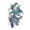
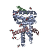
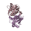
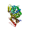
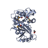
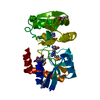
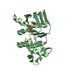
- PDB-2ehp: Crystal Structure of a Putative protein (AQ1627) from Aquifex aeolicus -
+ Open data

- Basic information
| Entry | Database: PDB / ID: 2ehp | ||||||
|---|---|---|---|---|---|---|---|
| Title | Crystal Structure of a Putative protein (AQ1627) from Aquifex aeolicus | ||||||
 Components Components | aq_1627 protein | ||||||
 Keywords Keywords | STRUCTURAL GENOMICS / UNKNOWN FUNCTION / Putative protein / Aquifex aeolicus / NPPSFA / National Project on Protein Structural and Functional Analyses / RIKEN Structural Genomics/Proteomics Initiative / RSGI | ||||||
| Function / homology | Alpha-D-Glucose-1,6-Bisphosphate; Chain A, domain 3 / Alpha-D-Glucose-1,6-Bisphosphate, subunit A, domain 3 / 3-Layer(aba) Sandwich / Alpha Beta / Uncharacterized protein aq_1627 Function and homology information Function and homology information | ||||||
| Biological species |   Aquifex aeolicus (bacteria) Aquifex aeolicus (bacteria) | ||||||
| Method |  X-RAY DIFFRACTION / SYNCHROTRON / MAD / Resolution: 1.3 Å X-RAY DIFFRACTION / SYNCHROTRON / MAD / Resolution: 1.3 Å | ||||||
 Authors Authors | Kumarevel, T.S. / Karthe, P. / Nakano, N. / Kuramitsu, S. / Yokoyama, S. / RIKEN Structural Genomics/Proteomics Initiative (RSGI) | ||||||
 Citation Citation | Journal: To be Published Title: Crystal Structure of a Putative protein (AQ1627) from Aquifex aeolicus Authors: Kumarevel, T.S. / Karthe, P. / Nakano, N. / Kuramitsu, S. / Yokoyama, S. | ||||||
| History |
|
- Structure visualization
| Structure viewer | Molecule: MolmilJmol/JSmol |
|---|
- Downloads & links
-Download
| PDBx/mmCIF format | 2ehp.cif.gz | 61.5 KB | Display | PDBx/mmCIF format |
|---|---|---|---|---|
| PDB format | pdb2ehp.ent.gz | 44.8 KB | Display | PDB format |
| PDBx/mmJSON format | 2ehp.json.gz | Tree view | PDBx/mmJSON format | |
| Others |  Other downloads Other downloads |
-Validation report
| Arichive directory | https://data.pdbj.org/pub/pdb/validation_reports/eh/2ehpftp://data.pdbj.org/pub/pdb/validation_reports/eh/2ehp | HTTPS FTP |
|---|
-Related structure data


-Links
 PDBj
PDBj- Assembly
| Deposited unit | 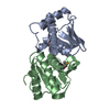
| ||||||||
|---|---|---|---|---|---|---|---|---|---|
| 1 |
| ||||||||
| Unit cell |
| ||||||||
| Components on special symmetry positions |
|
-Components
| #1: Protein | Mass: 13820.679 Da / Num. of mol.: 2 Source method: isolated from a genetically manipulated source Source: (gene. exp.) Aquifex aeolicus (bacteria) / Plasmid: pET-21a / Production host: #2: Water | ChemComp-HOH / |  Mass: 18.015 Da / Num. of mol.: 150 / Source method: isolated from a natural source / Formula: H2O Mass: 18.015 Da / Num. of mol.: 150 / Source method: isolated from a natural source / Formula: H2OHas protein modification | Y | |
|---|
-Experimental details
-Experiment
| Experiment | Method: X-RAY DIFFRACTION / Number of used crystals: 1 |
|---|
- Sample preparation
| Crystal | Density Matthews: 2.18 Å3/Da / Density % sol: 43.59 % |
|---|---|
| Crystal grow | Temperature: 293 K / Method: vapor diffusion, sitting drop / pH: 4.2 Details: 40% Ethanol, 5% PEG1000, Phosphate-Citrate, pH 4.2, VAPOR DIFFUSION, SITTING DROP, temperature 293K |
-Data collection
| Diffraction | Mean temperature: 180 K | ||||||||||||
|---|---|---|---|---|---|---|---|---|---|---|---|---|---|
| Diffraction source | Source: SYNCHROTRON / Site: SPring-8  / Beamline: BL26B2 / Wavelength: 0.97884, 0.9000, 0.97973 / Beamline: BL26B2 / Wavelength: 0.97884, 0.9000, 0.97973 | ||||||||||||
| Detector | Type: RIGAKU JUPITER 210 / Detector: CCD / Date: Nov 28, 2006 / Details: Si | ||||||||||||
| Radiation | Monochromator: Si / Protocol: MAD / Monochromatic (M) / Laue (L): M / Scattering type: x-ray | ||||||||||||
| Radiation wavelength |
| ||||||||||||
| Reflection | Resolution: 1.3→50 Å / Num. all: 51115 / Num. obs: 51115 / % possible obs: 89.3 % / Observed criterion σ(F): 2 / Observed criterion σ(I): 2 / Redundancy: 6.7 % / Biso Wilson estimate: 13.3 Å2 / Rmerge(I) obs: 0.034 | ||||||||||||
| Reflection shell | Resolution: 1.3→1.35 Å / Redundancy: 6.9 % / Rmerge(I) obs: 0.204 / Num. unique all: 4884 / % possible all: 85.7 |
- Processing
| Software |
| ||||||||||||||||||||||||||||||||||||
|---|---|---|---|---|---|---|---|---|---|---|---|---|---|---|---|---|---|---|---|---|---|---|---|---|---|---|---|---|---|---|---|---|---|---|---|---|---|
| Refinement | Method to determine structure: MAD / Resolution: 1.3→19.97 Å / Rfactor Rfree error: 0.005 / Data cutoff high absF: 1028214.05 / Data cutoff low absF: 0 / Isotropic thermal model: RESTRAINED / Cross valid method: THROUGHOUT / σ(F): 0
| ||||||||||||||||||||||||||||||||||||
| Solvent computation | Solvent model: FLAT MODEL / Bsol: 45.1226 Å2 / ksol: 0.347829 e/Å3 | ||||||||||||||||||||||||||||||||||||
| Displacement parameters | Biso mean: 15.7 Å2
| ||||||||||||||||||||||||||||||||||||
| Refine analyze |
| ||||||||||||||||||||||||||||||||||||
| Refinement step | Cycle: LAST / Resolution: 1.3→19.97 Å
| ||||||||||||||||||||||||||||||||||||
| Refine LS restraints |
| ||||||||||||||||||||||||||||||||||||
| LS refinement shell | Resolution: 1.3→1.38 Å / Rfactor Rfree error: 0.013 / Total num. of bins used: 6
| ||||||||||||||||||||||||||||||||||||
| Xplor file |
|
