 Movie
Movie Controller

[English] 日本語
 Yorodumi
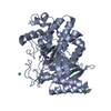
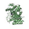
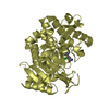
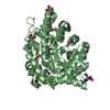
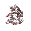
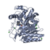
- PDB-2efu: The crystal structure of D-amino acid amidase from Ochrobactrum a... -
+ Open data

- Basic information
| Entry | Database: PDB / ID: 2efu | ||||||
|---|---|---|---|---|---|---|---|
| Title | The crystal structure of D-amino acid amidase from Ochrobactrum anthropi SV3 complexed with L-phenylalanine | ||||||
 Components Components | D-Amino acid amidase | ||||||
 Keywords Keywords | HYDROLASE / penicillin recognizing proteins / D-stereospecific / amidase / L-phenylalanine | ||||||
| Function / homology |  Function and homology information Function and homology information: / Beta-lactamase-related / Beta-lactamase / Beta-lactamase / DD-peptidase/beta-lactamase superfamily / Beta-lactamase/transpeptidase-like / 3-Layer(aba) Sandwich / Alpha Beta Similarity search - Domain/homology | ||||||
| Biological species |  Ochrobactrum anthropi (bacteria) Ochrobactrum anthropi (bacteria) | ||||||
| Method |  X-RAY DIFFRACTION / SYNCHROTRON / MOLECULAR REPLACEMENT / Resolution: 2.3 Å X-RAY DIFFRACTION / SYNCHROTRON / MOLECULAR REPLACEMENT / Resolution: 2.3 Å | ||||||
 Authors Authors | Okazaki, S. / Suzuki, A. / Mizushima, T. / Komeda, H. / Asano, Y. / Yamane, T. | ||||||
 Citation Citation | Journal: Acta Crystallogr.,Sect.D / Year: 2008 Title: Structures of D-amino-acid amidase complexed with L-phenylalanine and with L-phenylalanine amide: insight into the D-stereospecificity of D-amino-acid amidase from Ochrobactrum anthropi SV3. Authors: Okazaki, S. / Suzuki, A. / Mizushima, T. / Komeda, H. / Asano, Y. / Yamane, T. | ||||||
| History |
|
- Structure visualization
| Structure viewer | Molecule: MolmilJmol/JSmol |
|---|
- Downloads & links
-Download
| PDBx/mmCIF format | 2efu.cif.gz | 443.5 KB | Display | PDBx/mmCIF format |
|---|---|---|---|---|
| PDB format | pdb2efu.ent.gz | 359.7 KB | Display | PDB format |
| PDBx/mmJSON format | 2efu.json.gz | Tree view | PDBx/mmJSON format | |
| Others |  Other downloads Other downloads |
-Validation report
| Summary document | 2efu_validation.pdf.gz | 493.8 KB | Display | wwPDB validaton report |
|---|---|---|---|---|
| Full document | 2efu_full_validation.pdf.gz | 516.6 KB | Display | |
| Data in XML | 2efu_validation.xml.gz | 88.7 KB | Display | |
| Data in CIF | 2efu_validation.cif.gz | 125.8 KB | Display | |
| Arichive directory | https://data.pdbj.org/pub/pdb/validation_reports/ef/2efuftp://data.pdbj.org/pub/pdb/validation_reports/ef/2efu | HTTPS FTP |
-Related structure data
| Related structure data |  2efxC  2drwS C: citing same article ( S: Starting model for refinement |
|---|---|
| Similar structure data |


-Links
 PDBj
PDBj


- Assembly
| Deposited unit | 
| ||||||||
|---|---|---|---|---|---|---|---|---|---|
| 1 | 
| ||||||||
| 2 | 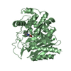
| ||||||||
| 3 | 
| ||||||||
| 4 | 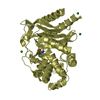
| ||||||||
| 5 | 
| ||||||||
| 6 | 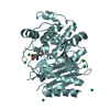
| ||||||||
| Unit cell |
|
-Components
| #1: Protein | Mass: 40127.191 Da / Num. of mol.: 6 Source method: isolated from a genetically manipulated source Source: (gene. exp.) Ochrobactrum anthropi (bacteria) / Strain: SV3 / Plasmid: pUC19 / Production host: #2: Chemical | ChemComp-BA /   Mass: 137.327 Da / Num. of mol.: 25 / Source method: obtained synthetically / Formula: Ba Mass: 137.327 Da / Num. of mol.: 25 / Source method: obtained synthetically / Formula: Ba#3: Chemical | ChemComp-PHE /   Type: L-peptide linking / Mass: 165.189 Da / Num. of mol.: 6 / Source method: obtained synthetically / Formula: C9H11NO2 Type: L-peptide linking / Mass: 165.189 Da / Num. of mol.: 6 / Source method: obtained synthetically / Formula: C9H11NO2#4: Water | ChemComp-HOH / |  Mass: 18.015 Da / Num. of mol.: 1407 / Source method: isolated from a natural source / Formula: H2O Mass: 18.015 Da / Num. of mol.: 1407 / Source method: isolated from a natural source / Formula: H2O |
|---|
-Experimental details
-Experiment
| Experiment | Method: X-RAY DIFFRACTION |
|---|
- Sample preparation
| Crystal | Density Matthews: 2.23 Å3/Da / Density % sol: 44.93 % |
|---|
-Data collection
| Diffraction | Mean temperature: 100 K |
|---|---|
| Diffraction source | Source: SYNCHROTRON / Site: SPring-8  / Beamline: BL38B1 / Wavelength: 1 Å / Beamline: BL38B1 / Wavelength: 1 Å |
| Detector | Type: RIGAKU JUPITER 210 / Detector: CCD / Date: Sep 24, 2005 |
| Radiation | Protocol: SINGLE WAVELENGTH / Monochromatic (M) / Laue (L): M / Scattering type: x-ray |
| Radiation wavelength | Wavelength: 1 Å / Relative weight: 1 |
| Reflection | Resolution: 2.3→50 Å / Num. obs: 93589 / % possible obs: 100 % / Observed criterion σ(I): 0 / Redundancy: 6.1 % / Rmerge(I) obs: 0.093 / Rsym value: 0.096 |
| Reflection shell | Resolution: 2.3→2.38 Å / Rmerge(I) obs: 0.359 / % possible all: 100 |
- Processing
| Software |
| ||||||||||||||||||||||||||||||||||||||||||||||||||||||||||||||||||||||||||||||||||||||||||||||||||||
|---|---|---|---|---|---|---|---|---|---|---|---|---|---|---|---|---|---|---|---|---|---|---|---|---|---|---|---|---|---|---|---|---|---|---|---|---|---|---|---|---|---|---|---|---|---|---|---|---|---|---|---|---|---|---|---|---|---|---|---|---|---|---|---|---|---|---|---|---|---|---|---|---|---|---|---|---|---|---|---|---|---|---|---|---|---|---|---|---|---|---|---|---|---|---|---|---|---|---|---|---|---|
| Refinement | Method to determine structure: MOLECULAR REPLACEMENT Starting model: DAA native structure (2DRW) Resolution: 2.3→47.67 Å / Cor.coef. Fo:Fc: 0.949 / Cor.coef. Fo:Fc free: 0.9 / SU B: 6.647 / SU ML: 0.165 / Cross valid method: THROUGHOUT / ESU R: 0.403 / ESU R Free: 0.247 / Stereochemistry target values: MAXIMUM LIKELIHOOD / Details: HYDROGENS HAVE BEEN ADDED IN THE RIDING POSITIONS
| ||||||||||||||||||||||||||||||||||||||||||||||||||||||||||||||||||||||||||||||||||||||||||||||||||||
| Solvent computation | Ion probe radii: 0.8 Å / Shrinkage radii: 0.8 Å / VDW probe radii: 1.2 Å / Solvent model: MASK | ||||||||||||||||||||||||||||||||||||||||||||||||||||||||||||||||||||||||||||||||||||||||||||||||||||
| Displacement parameters | Biso mean: 25.417 Å2
| ||||||||||||||||||||||||||||||||||||||||||||||||||||||||||||||||||||||||||||||||||||||||||||||||||||
| Refinement step | Cycle: LAST / Resolution: 2.3→47.67 Å
| ||||||||||||||||||||||||||||||||||||||||||||||||||||||||||||||||||||||||||||||||||||||||||||||||||||
| Refine LS restraints |
| ||||||||||||||||||||||||||||||||||||||||||||||||||||||||||||||||||||||||||||||||||||||||||||||||||||
| LS refinement shell | Resolution: 2.3→2.36 Å / Total num. of bins used: 20
|
