 Movie
Movie Controller

[English] 日本語
 Yorodumi
- PDB-2ebh: Crystal structures reveal a thiol-protease like catalytic triad i... -
+ Open data

- Basic information
| Entry | Database: PDB / ID: 2ebh | |||||||||
|---|---|---|---|---|---|---|---|---|---|---|
| Title | Crystal structures reveal a thiol-protease like catalytic triad in the C-terminal region of Pasteurella multocida toxin | |||||||||
 Components Components | Dermonecrotic toxin | |||||||||
 Keywords Keywords | TOXIN / Pasteurella multocida toxin / Cys1165Ser mutant / inactivated mutant | |||||||||
| Function / homology |  Function and homology information Function and homology informationsymbiont-mediated perturbation of host G protein-coupled receptor signal transduction pathway / glycerophospholipase activity / symbiont-mediated perturbation of host cell cycle progression / symbiont-mediated killing of host cell / phospholipid binding / toxin activity / host cell plasma membrane / extracellular region / cytoplasm Similarity search - Function | |||||||||
| Biological species |  Pasteurella multocida (bacteria) Pasteurella multocida (bacteria) | |||||||||
| Method |  X-RAY DIFFRACTION / SYNCHROTRON / MOLECULAR REPLACEMENT / Resolution: 2.4 Å X-RAY DIFFRACTION / SYNCHROTRON / MOLECULAR REPLACEMENT / Resolution: 2.4 Å | |||||||||
 Authors Authors | Kitadokoro, K. / Horiguchi, Y. / Kamitani, S. | |||||||||
 Citation Citation | Journal: Proc.Natl.Acad.Sci.Usa / Year: 2007 Title: Crystal structures reveal a thiol protease-like catalytic triad in the C-terminal region of Pasteurella multocida toxin Authors: Kitadokoro, K. / Kamitani, S. / Miyazawa, M. / Hanajima-Ozawa, M. / Fukui, A. / Miyake, M. / Horiguchi, Y. | |||||||||
| History |
|
- Structure visualization
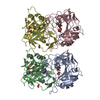
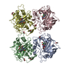
| Structure viewer | Molecule: MolmilJmol/JSmol |
|---|
- Downloads & links
-Download
| PDBx/mmCIF format | 2ebh.cif.gz | 163.8 KB | Display | PDBx/mmCIF format |
|---|---|---|---|---|
| PDB format | pdb2ebh.ent.gz | 125.1 KB | Display | PDB format |
| PDBx/mmJSON format | 2ebh.json.gz | Tree view | PDBx/mmJSON format | |
| Others |  Other downloads Other downloads |
-Validation report
| Arichive directory | https://data.pdbj.org/pub/pdb/validation_reports/eb/2ebhftp://data.pdbj.org/pub/pdb/validation_reports/eb/2ebh | HTTPS FTP |
|---|
-Related structure data
| Related structure data | 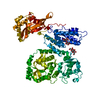 2ebfSC 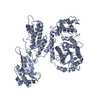 2ec5C S: Starting model for refinement C: citing same article ( |
|---|---|
| Similar structure data |


-Links
 PDBj
PDBj
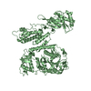
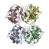
- Assembly
| Deposited unit | 
| ||||||||
|---|---|---|---|---|---|---|---|---|---|
| 1 |
| ||||||||
| Unit cell |
| ||||||||
| Components on special symmetry positions |
| ||||||||
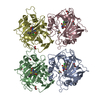
| Details | The molecule exist as a monomer |
-Components
| #1: Protein | Mass: 84281.539 Da / Num. of mol.: 1 / Fragment: C-terminal region, residues 569-1285 / Mutation: C1165S Source method: isolated from a genetically manipulated source Source: (gene. exp.) Pasteurella multocida (bacteria) / Strain: G-7 / Gene: toxA / Plasmid: pPROEX-1, pPROEX-1-C-PMT / Species (production host): Escherichia coli / Production host: | ||
|---|---|---|---|
| #2: Polysaccharide |   Source method: isolated from a genetically manipulated source Details: oligosaccharide with reducing-end-to-reducing-end glycosidic bond References: trehalose #3: Water | ChemComp-HOH / |  Mass: 18.015 Da / Num. of mol.: 371 / Source method: isolated from a natural source / Formula: H2O Mass: 18.015 Da / Num. of mol.: 371 / Source method: isolated from a natural source / Formula: H2O |
-Experimental details
-Experiment
| Experiment | Method: X-RAY DIFFRACTION / Number of used crystals: 1 |
|---|
- Sample preparation
| Crystal | Density Matthews: 3.73 Å3/Da / Density % sol: 67.01 % |
|---|---|
| Crystal grow | Temperature: 293 K / Method: vapor diffusion, sitting drop / pH: 6.5 Details: 1.0M ammonium citrate, 0.1M MES, pH 6.5, VAPOR DIFFUSION, SITTING DROP, temperature 293K |
-Data collection
| Diffraction | Mean temperature: 100 K |
|---|---|
| Diffraction source | Source: SYNCHROTRON / Site: SPring-8  / Beamline: BL44XU / Wavelength: 0.9 Å / Beamline: BL44XU / Wavelength: 0.9 Å |
| Detector | Type: MACSCIENCE / Detector: IMAGE PLATE / Date: May 22, 2006 / Details: mirrors |
| Radiation | Protocol: SINGLE WAVELENGTH / Monochromatic (M) / Laue (L): M / Scattering type: x-ray |
| Radiation wavelength | Wavelength: 0.9 Å / Relative weight: 1 |
| Reflection | Resolution: 2.4→50 Å / Num. all: 48566 / Num. obs: 47787 / % possible obs: 99.1 % / Redundancy: 3.8 % / Rmerge(I) obs: 0.094 |
| Reflection shell | Resolution: 2.4→2.49 Å / Redundancy: 3.5 % / Rmerge(I) obs: 0.403 / Num. unique all: 4466 / % possible all: 93.5 |
- Processing
| Software |
| ||||||||||||||||||||||||||||||||||||||||||||||||||||||||||||||||||||||||||||||||||||||||||
|---|---|---|---|---|---|---|---|---|---|---|---|---|---|---|---|---|---|---|---|---|---|---|---|---|---|---|---|---|---|---|---|---|---|---|---|---|---|---|---|---|---|---|---|---|---|---|---|---|---|---|---|---|---|---|---|---|---|---|---|---|---|---|---|---|---|---|---|---|---|---|---|---|---|---|---|---|---|---|---|---|---|---|---|---|---|---|---|---|---|---|---|
| Refinement | Method to determine structure: MOLECULAR REPLACEMENT Starting model: PDB ENTRY 2EBF Resolution: 2.4→43.69 Å / Cor.coef. Fo:Fc: 0.939 / Cor.coef. Fo:Fc free: 0.919 / SU B: 7.886 / SU ML: 0.177 / Cross valid method: THROUGHOUT / σ(F): 0 / σ(I): 0.403 / ESU R: 0.28 / ESU R Free: 0.224 / Stereochemistry target values: MAXIMUM LIKELIHOOD / Details: HYDROGENS HAVE BEEN ADDED IN THE RIDING POSITIONS
| ||||||||||||||||||||||||||||||||||||||||||||||||||||||||||||||||||||||||||||||||||||||||||
| Solvent computation | Ion probe radii: 0.8 Å / Shrinkage radii: 0.8 Å / VDW probe radii: 1.2 Å / Solvent model: MASK | ||||||||||||||||||||||||||||||||||||||||||||||||||||||||||||||||||||||||||||||||||||||||||
| Displacement parameters | Biso mean: 43.807 Å2
| ||||||||||||||||||||||||||||||||||||||||||||||||||||||||||||||||||||||||||||||||||||||||||
| Refinement step | Cycle: LAST / Resolution: 2.4→43.69 Å
| ||||||||||||||||||||||||||||||||||||||||||||||||||||||||||||||||||||||||||||||||||||||||||
| Refine LS restraints |
| ||||||||||||||||||||||||||||||||||||||||||||||||||||||||||||||||||||||||||||||||||||||||||
| LS refinement shell | Resolution: 2.4→2.462 Å / Total num. of bins used: 20
|
