 Movie
Movie Controller

[English] 日本語
 Yorodumi
- PDB-2e4o: X-ray Crystal Structure of Aristolochene Synthase from Aspergillu... -
+ Open data

- Basic information
| Entry | Database: PDB / ID: 2e4o | ||||||
|---|---|---|---|---|---|---|---|
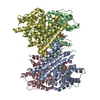
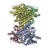
| Title | X-ray Crystal Structure of Aristolochene Synthase from Aspergillus terreus and the Evolution of Templates for the Cyclization of Farnesyl Diphosphate | ||||||
 Components Components | Aristolochene synthase | ||||||
 Keywords Keywords | LYASE / sesquiterpene cyclase / farnesyl diphosphate | ||||||
| Function / homology |  Function and homology information Function and homology informationaristolochene synthase / aristolochene synthase activity / isoprenoid biosynthetic process / metal ion binding Similarity search - Function | ||||||
| Biological species |  | ||||||
| Method |  X-RAY DIFFRACTION / SYNCHROTRON / MOLECULAR REPLACEMENT / Resolution: 2.2 Å X-RAY DIFFRACTION / SYNCHROTRON / MOLECULAR REPLACEMENT / Resolution: 2.2 Å | ||||||
 Authors Authors | Shishova, E.Y. / Di Costanzo, L. / Cane, D.E. / Christianson, D.W. | ||||||
 Citation Citation | Journal: Biochemistry / Year: 2007 Title: X-ray crystal structure of aristolochene synthase from Aspergillus terreus and evolution of templates for the cyclization of farnesyl diphosphate. Authors: Shishova, E.Y. / Di Costanzo, L. / Cane, D.E. / Christianson, D.W. | ||||||
| History |
|
- Structure visualization
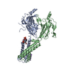
| Structure viewer | Molecule: MolmilJmol/JSmol |
|---|
- Downloads & links
-Download
| PDBx/mmCIF format | 2e4o.cif.gz | 246.1 KB | Display | PDBx/mmCIF format |
|---|---|---|---|---|
| PDB format | pdb2e4o.ent.gz | 200 KB | Display | PDB format |
| PDBx/mmJSON format | 2e4o.json.gz | Tree view | PDBx/mmJSON format | |
| Others |  Other downloads Other downloads |
-Validation report
| Arichive directory | https://data.pdbj.org/pub/pdb/validation_reports/e4/2e4oftp://data.pdbj.org/pub/pdb/validation_reports/e4/2e4o | HTTPS FTP |
|---|
-Related structure data
| Related structure data | 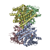 2oa6C 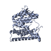 1di1S S: Starting model for refinement C: citing same article ( |
|---|---|
| Similar structure data |


-Links
 PDBj
PDBj


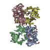
- Assembly
| Deposited unit | 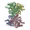
| ||||||||
|---|---|---|---|---|---|---|---|---|---|
| 1 |
| ||||||||
| Unit cell |
|
-Components
| #1: Protein | Mass: 36523.715 Da / Num. of mol.: 4 Source method: isolated from a genetically manipulated source Source: (gene. exp.)  #2: Chemical | ChemComp-BME /   Mass: 78.133 Da / Num. of mol.: 5 / Source method: obtained synthetically / Formula: C2H6OS Mass: 78.133 Da / Num. of mol.: 5 / Source method: obtained synthetically / Formula: C2H6OS#3: Chemical |   Mass: 35.453 Da / Num. of mol.: 2 / Source method: obtained synthetically / Formula: Cl Mass: 35.453 Da / Num. of mol.: 2 / Source method: obtained synthetically / Formula: Cl#4: Chemical | ChemComp-MES / |   Mass: 195.237 Da / Num. of mol.: 1 / Source method: obtained synthetically / Formula: C6H13NO4S / Comment: pH buffer*YM Mass: 195.237 Da / Num. of mol.: 1 / Source method: obtained synthetically / Formula: C6H13NO4S / Comment: pH buffer*YM#5: Water | ChemComp-HOH / |  Mass: 18.015 Da / Num. of mol.: 191 / Source method: isolated from a natural source / Formula: H2O Mass: 18.015 Da / Num. of mol.: 191 / Source method: isolated from a natural source / Formula: H2O |
|---|
-Experimental details
-Experiment
| Experiment | Method: X-RAY DIFFRACTION / Number of used crystals: 1 |
|---|
- Sample preparation
| Crystal | Density Matthews: 2.56 Å3/Da / Density % sol: 51.88 % |
|---|---|
| Crystal grow | Temperature: 277 K / Method: vapor diffusion, hanging drop / pH: 8.5 Details: 100mM Tris, 18% PEG 6000, 0.5M NaCl, pH 8.5, VAPOR DIFFUSION, HANGING DROP, temperature 277K |
-Data collection
| Diffraction | Mean temperature: 100 K |
|---|---|
| Diffraction source | Source: SYNCHROTRON / Site: NSLS  / Beamline: X29A / Wavelength: 1 Å / Beamline: X29A / Wavelength: 1 Å |
| Detector | Type: ADSC QUANTUM 315 / Detector: CCD / Date: Jun 6, 2005 |
| Radiation | Protocol: SINGLE WAVELENGTH / Monochromatic (M) / Laue (L): M / Scattering type: x-ray |
| Radiation wavelength | Wavelength: 1 Å / Relative weight: 1 |
| Reflection | Resolution: 2.2→50 Å / Num. all: 73132 / Num. obs: 72129 / % possible obs: 96.2 % / Observed criterion σ(I): 2.3 / Redundancy: 2.9 % / Biso Wilson estimate: 36.7 Å2 / Rmerge(I) obs: 0.084 / Net I/σ(I): 19 |
| Reflection shell | Resolution: 2.2→2.28 Å / Redundancy: 2.7 % / Rmerge(I) obs: 0.534 / Mean I/σ(I) obs: 2.26 / Num. unique all: 6773 / % possible all: 89.1 |
- Processing
| Software |
| ||||||||||||||||||||
|---|---|---|---|---|---|---|---|---|---|---|---|---|---|---|---|---|---|---|---|---|---|
| Refinement | Method to determine structure: MOLECULAR REPLACEMENT Starting model: 1DI1 Resolution: 2.2→50 Å / Cross valid method: THROUGHOUT / Stereochemistry target values: cns library
| ||||||||||||||||||||
| Refinement step | Cycle: LAST / Resolution: 2.2→50 Å
| ||||||||||||||||||||
| Refine LS restraints |
|
