 Movie
Movie Controller


+ Open data

- Basic information
| Entry | Database: PDB / ID: 2dub | ||||||
|---|---|---|---|---|---|---|---|
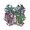
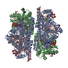
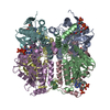
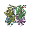
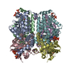
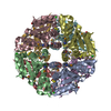
| Title | ENOYL-COA HYDRATASE COMPLEXED WITH OCTANOYL-COA | ||||||
 Components Components | 2-ENOYL-COA HYDRATASE | ||||||
 Keywords Keywords | LYASE / HYDRATASE / B-OXIDATION / FATTY ACID DEGRADATION / COA / LIGAND BINDING | ||||||
| Function / homology |  Function and homology information Function and homology informationBeta oxidation of lauroyl-CoA to decanoyl-CoA-CoA / Beta oxidation of decanoyl-CoA to octanoyl-CoA-CoA / Beta oxidation of octanoyl-CoA to hexanoyl-CoA / Beta oxidation of hexanoyl-CoA to butanoyl-CoA / Beta oxidation of butanoyl-CoA to acetyl-CoA / 3-hydroxypropionyl-CoA dehydratase activity / (2E)-butenoyl-CoA hydratase activity / Branched-chain amino acid catabolism / Delta3-Delta2-enoyl-CoA isomerase / delta(3)-delta(2)-enoyl-CoA isomerase activity ...Beta oxidation of lauroyl-CoA to decanoyl-CoA-CoA / Beta oxidation of decanoyl-CoA to octanoyl-CoA-CoA / Beta oxidation of octanoyl-CoA to hexanoyl-CoA / Beta oxidation of hexanoyl-CoA to butanoyl-CoA / Beta oxidation of butanoyl-CoA to acetyl-CoA / 3-hydroxypropionyl-CoA dehydratase activity / (2E)-butenoyl-CoA hydratase activity / Branched-chain amino acid catabolism / Delta3-Delta2-enoyl-CoA isomerase / delta(3)-delta(2)-enoyl-CoA isomerase activity / enoyl-CoA hydratase / enoyl-CoA hydratase activity / fatty acid beta-oxidation / mitochondrial matrix / mitochondrion Similarity search - Function | ||||||
| Biological species |  | ||||||
| Method |  X-RAY DIFFRACTION / SYNCHROTRON / MOLECULAR REPLACEMENT / Resolution: 2.4 Å X-RAY DIFFRACTION / SYNCHROTRON / MOLECULAR REPLACEMENT / Resolution: 2.4 Å | ||||||
 Authors Authors | Engel, C.K. / Wierenga, R.K. | ||||||
 Citation Citation | Journal: J.Mol.Biol. / Year: 1998 Title: The crystal structure of enoyl-CoA hydratase complexed with octanoyl-CoA reveals the structural adaptations required for binding of a long chain fatty acid-CoA molecule. Authors: Engel, C.K. / Kiema, T.R. / Hiltunen, J.K. / Wierenga, R.K. #1: Journal: Embo J. / Year: 1996Title: Crystal Structure of Enoyl-Coenzyme a (Coa) Hydratase at 2.5 Angstroms Resolution: A Spiral Fold Defines the Coa-Binding Pocket Authors: Engel, C.K. / Mathieu, M. / Zeelen, J.P. / Hiltunen, J.K. / Wierenga, R.K. | ||||||
| History |
|
- Structure visualization
| Structure viewer | Molecule: MolmilJmol/JSmol |
|---|
- Downloads & links
-Download
| PDBx/mmCIF format | 2dub.cif.gz | 306.3 KB | Display | PDBx/mmCIF format |
|---|---|---|---|---|
| PDB format | pdb2dub.ent.gz | 249.9 KB | Display | PDB format |
| PDBx/mmJSON format | 2dub.json.gz | Tree view | PDBx/mmJSON format | |
| Others |  Other downloads Other downloads |
-Validation report
| Arichive directory | https://data.pdbj.org/pub/pdb/validation_reports/du/2dubftp://data.pdbj.org/pub/pdb/validation_reports/du/2dub | HTTPS FTP |
|---|
-Related structure data
| Related structure data |  1dubS S: Starting model for refinement |
|---|---|
| Similar structure data |


-Links
 PDBj
PDBj- Assembly
| Deposited unit | 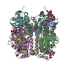
| ||||||||||||||||||||||||
|---|---|---|---|---|---|---|---|---|---|---|---|---|---|---|---|---|---|---|---|---|---|---|---|---|---|
| 1 |
| ||||||||||||||||||||||||
| 2 | 
| ||||||||||||||||||||||||
| Unit cell |
| ||||||||||||||||||||||||
| Noncrystallographic symmetry (NCS) | NCS oper:
|
-Components
| #1: Protein | Mass: 28321.514 Da / Num. of mol.: 6 / Source method: isolated from a natural source / Details: COCRYSTALLIZED WITH OCTANOYL-COA / Source: (natural) #2: Chemical | ChemComp-CO8 /   Mass: 893.730 Da / Num. of mol.: 4 / Source method: obtained synthetically / Formula: C29H50N7O17P3S Mass: 893.730 Da / Num. of mol.: 4 / Source method: obtained synthetically / Formula: C29H50N7O17P3S#3: Water | ChemComp-HOH / |  Mass: 18.015 Da / Num. of mol.: 436 / Source method: isolated from a natural source / Formula: H2O Mass: 18.015 Da / Num. of mol.: 436 / Source method: isolated from a natural source / Formula: H2OHas protein modification | N | |
|---|
-Experimental details
-Experiment
| Experiment | Method: X-RAY DIFFRACTION / Number of used crystals: 1 |
|---|
- Sample preparation
| Crystal | Density Matthews: 2.6 Å3/Da / Density % sol: 53 % Description: MOLECULAR REPLACEMENT BY RIGID-BODY REFINEMENT IN X-PLOR | ||||||||||||||||||||||||||||||||||||||||||||||||||||||||||||||||||||||||||||||||||||||||||
|---|---|---|---|---|---|---|---|---|---|---|---|---|---|---|---|---|---|---|---|---|---|---|---|---|---|---|---|---|---|---|---|---|---|---|---|---|---|---|---|---|---|---|---|---|---|---|---|---|---|---|---|---|---|---|---|---|---|---|---|---|---|---|---|---|---|---|---|---|---|---|---|---|---|---|---|---|---|---|---|---|---|---|---|---|---|---|---|---|---|---|---|
| Crystal grow | pH: 7.5 Details: PROTEIN SOLUTION: 4.5 MG/ML PROTEIN IN 20 MM POTASSIUM PHOSPHATE BUFFER, PH 7.2, 3 MM EDTA, 1 MM OCTANOYL-COA RESERVOIR SOLUTION: 2.2 M AMMONIUM SULFATE, 200 MM TRIETHANOLAMINE PH 7.5, 1MM ...Details: PROTEIN SOLUTION: 4.5 MG/ML PROTEIN IN 20 MM POTASSIUM PHOSPHATE BUFFER, PH 7.2, 3 MM EDTA, 1 MM OCTANOYL-COA RESERVOIR SOLUTION: 2.2 M AMMONIUM SULFATE, 200 MM TRIETHANOLAMINE PH 7.5, 1MM DTT, 1MM NAN3, 1MM EDTA, 5% (V/V) N-OCTANOYL CRYSTALS GREW BY MIXING 1 MICROLITER PROTEIN SOLUTION WITH 1 MICROLITER RESERVOIR SOLUTION PH range: 7.2-7.5 | ||||||||||||||||||||||||||||||||||||||||||||||||||||||||||||||||||||||||||||||||||||||||||
| Crystal | *PLUS | ||||||||||||||||||||||||||||||||||||||||||||||||||||||||||||||||||||||||||||||||||||||||||
| Crystal grow | *PLUS pH: 7.2 / Method: vapor diffusion, hanging drop | ||||||||||||||||||||||||||||||||||||||||||||||||||||||||||||||||||||||||||||||||||||||||||
| Components of the solutions | *PLUS
|
-Data collection
| Diffraction | Mean temperature: 100 K |
|---|---|
| Diffraction source | Source: SYNCHROTRON / Site: EMBL/DESY, HAMBURG  / Beamline: X11 / Wavelength: 0.9065 / Beamline: X11 / Wavelength: 0.9065 |
| Detector | Type: MARRESEARCH / Detector: IMAGE PLATE / Date: Oct 1, 1996 / Details: COLLIMATOR |
| Radiation | Monochromator: CRYSTAL / Monochromatic (M) / Laue (L): M / Scattering type: x-ray |
| Radiation wavelength | Wavelength: 0.9065 Å / Relative weight: 1 |
| Reflection | Resolution: 2.4→20 Å / Num. obs: 68481 / % possible obs: 99.6 % / Observed criterion σ(I): 4 / Redundancy: 3.95 % / Rsym value: 0.075 / Net I/σ(I): 7.7 |
| Reflection shell | Resolution: 2.4→2.44 Å / Mean I/σ(I) obs: 4.1 / Rsym value: 0.316 / % possible all: 96 |
| Reflection | *PLUS Num. obs: 69852 / Num. measured all: 275878 / Rmerge(I) obs: 0.075 |
| Reflection shell | *PLUS % possible obs: 96 % / Num. unique obs: 3307 / Rmerge(I) obs: 0.316 |
- Processing
| Software |
| ||||||||||||||||||||
|---|---|---|---|---|---|---|---|---|---|---|---|---|---|---|---|---|---|---|---|---|---|
| Refinement | Method to determine structure: MOLECULAR REPLACEMENT Starting model: PDB ENTRY 1DUB Resolution: 2.4→20 Å / Cross valid method: THROUGHOUT / σ(F): 0 Details: THE STRUCTURE WAS REFINED USING THE PROGRAMS X-PLOR AND REFMAC. AFTER STRUCTURE DETERMINATION BY RIGID-BODY REFINEMENT (MODEL: PDB ENTRY 1DUB, WITHOUT LIGAND AND WATER MOLECULES), A SLOW ...Details: THE STRUCTURE WAS REFINED USING THE PROGRAMS X-PLOR AND REFMAC. AFTER STRUCTURE DETERMINATION BY RIGID-BODY REFINEMENT (MODEL: PDB ENTRY 1DUB, WITHOUT LIGAND AND WATER MOLECULES), A SLOW COOL SIMULATED ANNEALING PROTOCOL WAS RUN IN X-PLOR. SUBSEQUENT REFINEMENT WAS DONE WITH REFMAC. FOR BOTH PROGRAMS THE SAME REFLECTIONS WERE USED FOR CROSS VALIDATION. AN X-PLOR BULK SOLVENT CORRECTION WAS USED. AT THE END OF REFINEMENT A POSITIONAL REFINEMENT WAS RUN IN X-PLOR.
| ||||||||||||||||||||
| Refinement step | Cycle: LAST / Resolution: 2.4→20 Å
| ||||||||||||||||||||
| Software | *PLUS Name: REFMAC / Classification: refinement | ||||||||||||||||||||
| Refinement | *PLUS Num. reflection all: 68481 / Rfactor obs: 0.204 / Rfactor Rfree: 0.26 | ||||||||||||||||||||
| Solvent computation | *PLUS | ||||||||||||||||||||
| Displacement parameters | *PLUS | ||||||||||||||||||||
| Refine LS restraints | *PLUS
|
