 Movie
Movie Controller

[English] 日本語
 Yorodumi
- PDB-2dtt: Crystal structure of 6-pyruvoyl tetrahydrobiopterin synthase from... -
+ Open data

- Basic information
| Entry | Database: PDB / ID: 2dtt | ||||||
|---|---|---|---|---|---|---|---|
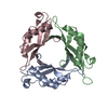
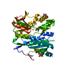
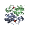
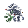
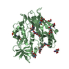
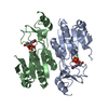
| Title | Crystal structure of 6-pyruvoyl tetrahydrobiopterin synthase from Pyrococcus horikoshii OT3 complexed with (1'R,2'S)-biopterin | ||||||
 Components Components | Hypothetical protein PH0634 | ||||||
 Keywords Keywords | LYASE / 6-pyruvoyl tetrahydrobiopterin synthase (PTPS) / Biopterin / Structural Genomics / NPPSFA / National Project on Protein Structural and Functional Analyses / RIKEN Structural Genomics/Proteomics Initiative / RSGI | ||||||
| Function / homology |  Function and homology information Function and homology information | ||||||
| Biological species |   Pyrococcus horikoshii (archaea) Pyrococcus horikoshii (archaea) | ||||||
| Method |  X-RAY DIFFRACTION / SYNCHROTRON / MOLECULAR REPLACEMENT / Resolution: 2.2 Å X-RAY DIFFRACTION / SYNCHROTRON / MOLECULAR REPLACEMENT / Resolution: 2.2 Å | ||||||
 Authors Authors | Bagautdinov, B. / Kunishima, N. / RIKEN Structural Genomics/Proteomics Initiative (RSGI) | ||||||
 Citation Citation | Journal: To be Published Title: Crystal structure of 6-pyruvoyl tetrahydrobiopterin synthase from Pyrococcus horikoshii OT3 Authors: Bagautdinov, B. / Kunishima, N. | ||||||
| History |
|
- Structure visualization
| Structure viewer | Molecule: MolmilJmol/JSmol |
|---|
- Downloads & links
-Download
| PDBx/mmCIF format | 2dtt.cif.gz | 154.8 KB | Display | PDBx/mmCIF format |
|---|---|---|---|---|
| PDB format | pdb2dtt.ent.gz | 123.2 KB | Display | PDB format |
| PDBx/mmJSON format | 2dtt.json.gz | Tree view | PDBx/mmJSON format | |
| Others |  Other downloads Other downloads |
-Validation report
| Arichive directory | https://data.pdbj.org/pub/pdb/validation_reports/dt/2dttftp://data.pdbj.org/pub/pdb/validation_reports/dt/2dtt | HTTPS FTP |
|---|
-Related structure data
| Related structure data |  2dj6SC S: Starting model for refinement C: citing same article ( |
|---|---|
| Similar structure data | |
| Other databases |


-Links
 PDBj
PDBj


- Assembly
| Deposited unit | 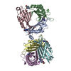
| ||||||||
|---|---|---|---|---|---|---|---|---|---|
| 1 | 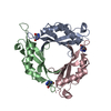
| ||||||||
| 2 | 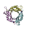
| ||||||||
| Unit cell |
| ||||||||
| Details | Biological assembly is a trimer. In the assymmetric unit subunits (A,B,C) and (D,E,F) represent biological units |
-Components
| #1: Protein | Mass: 13532.464 Da / Num. of mol.: 6 Source method: isolated from a genetically manipulated source Source: (gene. exp.) Pyrococcus horikoshii (archaea) / Strain: OT3 / Gene: PH0634 / Plasmid: pET 11a / Production host:  #2: Chemical | ChemComp-H4B /   Mass: 241.247 Da / Num. of mol.: 6 / Source method: obtained synthetically / Formula: C9H15N5O3 Mass: 241.247 Da / Num. of mol.: 6 / Source method: obtained synthetically / Formula: C9H15N5O3#3: Water | ChemComp-HOH / |  Mass: 18.015 Da / Num. of mol.: 405 / Source method: isolated from a natural source / Formula: H2O Mass: 18.015 Da / Num. of mol.: 405 / Source method: isolated from a natural source / Formula: H2O |
|---|
-Experimental details
-Experiment
| Experiment | Method: X-RAY DIFFRACTION / Number of used crystals: 1 |
|---|
- Sample preparation
| Crystal | Density Matthews: 2.23 Å3/Da / Density % sol: 44.77 % |
|---|---|
| Crystal grow | Temperature: 291 K / Method: microbathch / pH: 6.25 Details: 16.5% PEG 20000, 0.1M Acetate, NAOH pH6.25, 3mM Biopterin, microbathch, temperature 291K |
-Data collection
| Diffraction | Mean temperature: 100 K |
|---|---|
| Diffraction source | Source: SYNCHROTRON / Site: SPring-8  / Beamline: BL26B1 / Wavelength: 1 Å / Beamline: BL26B1 / Wavelength: 1 Å |
| Detector | Type: MARRESEARCH / Detector: CCD / Date: May 21, 2006 / Details: mirrors |
| Radiation | Monochromator: GRAPHITE / Protocol: SINGLE WAVELENGTH / Monochromatic (M) / Laue (L): M / Scattering type: x-ray |
| Radiation wavelength | Wavelength: 1 Å / Relative weight: 1 |
| Reflection | Resolution: 2.2→50 Å / Num. all: 36076 / Num. obs: 32611 / % possible obs: 90.4 % / Observed criterion σ(F): 0 / Observed criterion σ(I): 0 / Redundancy: 3 % / Biso Wilson estimate: 32.7 Å2 / Rmerge(I) obs: 0.072 / Rsym value: 0.067 / Net I/σ(I): 8.3 |
| Reflection shell | Resolution: 2.2→2.28 Å / Redundancy: 3 % / Rmerge(I) obs: 0.229 / Mean I/σ(I) obs: 3.1 / Num. unique all: 2906 / Rsym value: 0.225 / % possible all: 81.4 |
- Processing
| Software |
| |||||||||||||||||||||||||
|---|---|---|---|---|---|---|---|---|---|---|---|---|---|---|---|---|---|---|---|---|---|---|---|---|---|---|
| Refinement | Method to determine structure: MOLECULAR REPLACEMENT Starting model: 2DJ6 Resolution: 2.2→35.68 Å / Isotropic thermal model: Overall / Cross valid method: THROUGHOUT / σ(F): 0 / σ(I): 0 / Stereochemistry target values: Engh & Huber
| |||||||||||||||||||||||||
| Displacement parameters | Biso mean: 38.8 Å2
| |||||||||||||||||||||||||
| Refine analyze |
| |||||||||||||||||||||||||
| Refinement step | Cycle: LAST / Resolution: 2.2→35.68 Å
| |||||||||||||||||||||||||
| Refine LS restraints |
| |||||||||||||||||||||||||
| LS refinement shell | Resolution: 2.2→2.28 Å / Rfactor Rfree error: 0.024
|
