 Movie
Movie Controller

[English] 日本語
 Yorodumi
- PDB-2dr5: Complex structure of CCA adding enzyme with mini-helix lacking CCA -
+ Open data

- Basic information
| Entry | Database: PDB / ID: 2dr5 | ||||||
|---|---|---|---|---|---|---|---|
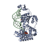
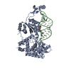
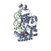
| Title | Complex structure of CCA adding enzyme with mini-helix lacking CCA | ||||||
 Components Components |
| ||||||
 Keywords Keywords | TRANSFERASE/RNA / protein-rna complex / TRANSFERASE-RNA COMPLEX | ||||||
| Function / homology |  Function and homology information Function and homology informationCCA tRNA nucleotidyltransferase / tRNA surveillance / CCA tRNA nucleotidyltransferase activity / CCACCA tRNA nucleotidyltransferase activity / tRNA 3'-terminal CCA addition / RNA repair / tRNA binding / magnesium ion binding / ATP binding Similarity search - Function | ||||||
| Biological species |   Archaeoglobus fulgidus (archaea) Archaeoglobus fulgidus (archaea) | ||||||
| Method |  X-RAY DIFFRACTION / SYNCHROTRON / MOLECULAR REPLACEMENT / Resolution: 2.8 Å X-RAY DIFFRACTION / SYNCHROTRON / MOLECULAR REPLACEMENT / Resolution: 2.8 Å | ||||||
 Authors Authors | Tomita, K. / Ishitani, R. / Fukai, S. / Nureki, O. | ||||||
 Citation Citation | Journal: Nature / Year: 2006 Title: Complete crystallographic analysis of the dynamics of CCA sequence addition Authors: Tomita, K. / Ishitani, R. / Fukai, S. / Nureki, O. | ||||||
| History |
|
- Structure visualization
| Structure viewer | Molecule: MolmilJmol/JSmol |
|---|
- Downloads & links
-Download
| PDBx/mmCIF format | 2dr5.cif.gz | 123.7 KB | Display | PDBx/mmCIF format |
|---|---|---|---|---|
| PDB format | pdb2dr5.ent.gz | 92.6 KB | Display | PDB format |
| PDBx/mmJSON format | 2dr5.json.gz | Tree view | PDBx/mmJSON format | |
| Others |  Other downloads Other downloads |
-Validation report
| Arichive directory | https://data.pdbj.org/pub/pdb/validation_reports/dr/2dr5ftp://data.pdbj.org/pub/pdb/validation_reports/dr/2dr5 | HTTPS FTP |
|---|
-Related structure data
| Related structure data |  2dr7C 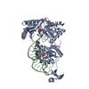 2dr8C  2dr9C  2draC  2drbC 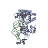 2dviC  1uetS C: citing same article ( S: Starting model for refinement |
|---|---|
| Similar structure data |


-Links
 PDBj
PDBj


- Assembly
| Deposited unit | 
| ||||||||
|---|---|---|---|---|---|---|---|---|---|
| 1 |
| ||||||||
| Unit cell |
|
-Components
| #1: RNA chain | Mass: 10294.140 Da / Num. of mol.: 1 / Source method: obtained synthetically | ||
|---|---|---|---|
| #2: Protein | Mass: 51469.137 Da / Num. of mol.: 1 Source method: isolated from a genetically manipulated source Source: (gene. exp.) Archaeoglobus fulgidus (archaea) / Production host:  | ||
| #3: Chemical |   Mass: 96.063 Da / Num. of mol.: 2 / Source method: obtained synthetically / Formula: SO4 Mass: 96.063 Da / Num. of mol.: 2 / Source method: obtained synthetically / Formula: SO4#4: Water | ChemComp-HOH / |  Mass: 18.015 Da / Num. of mol.: 103 / Source method: isolated from a natural source / Formula: H2O Mass: 18.015 Da / Num. of mol.: 103 / Source method: isolated from a natural source / Formula: H2O |
-Experimental details
-Experiment
| Experiment | Method: X-RAY DIFFRACTION / Number of used crystals: 1 |
|---|
- Sample preparation
| Crystal | Density Matthews: 3.02 Å3/Da / Density % sol: 59.26 % | ||||||||||||||||||||||||||||||||||||||||||||
|---|---|---|---|---|---|---|---|---|---|---|---|---|---|---|---|---|---|---|---|---|---|---|---|---|---|---|---|---|---|---|---|---|---|---|---|---|---|---|---|---|---|---|---|---|---|
| Crystal grow | Temperature: 293 K / Method: vapor diffusion / pH: 7.5 Details: 50mM HEPES, 20% PEG 3550, 0.2M Tri-lithium citrate, 80mM NH4SO4, pH 7.5, VAPOR DIFFUSION, temperature 293K | ||||||||||||||||||||||||||||||||||||||||||||
| Components of the solutions |
|
-Data collection
| Diffraction | Mean temperature: 100 K |
|---|---|
| Diffraction source | Source: SYNCHROTRON / Site: SPring-8  / Beamline: BL41XU / Wavelength: 1 Å / Beamline: BL41XU / Wavelength: 1 Å |
| Detector | Type: ADSC QUANTUM 315 / Detector: CCD / Date: May 17, 2005 |
| Radiation | Monochromator: Si 111 CHANNEL / Protocol: SINGLE WAVELENGTH / Monochromatic (M) / Laue (L): M / Scattering type: x-ray |
| Radiation wavelength | Wavelength: 1 Å / Relative weight: 1 |
| Reflection | Resolution: 2.8→50 Å / Num. all: 19376 / Num. obs: 19376 / % possible obs: 100 % / Observed criterion σ(F): 0 / Observed criterion σ(I): 0 / Biso Wilson estimate: 80 Å2 |
| Reflection shell | Resolution: 2.8→2.9 Å / % possible all: 97.7 |
- Processing
| Software |
| ||||||||||||||||||||||||||||||||||||
|---|---|---|---|---|---|---|---|---|---|---|---|---|---|---|---|---|---|---|---|---|---|---|---|---|---|---|---|---|---|---|---|---|---|---|---|---|---|
| Refinement | Method to determine structure: MOLECULAR REPLACEMENT Starting model: 1UET Resolution: 2.8→10 Å / Rfactor Rfree error: 0.01 / Data cutoff high absF: 2888737.69 / Data cutoff low absF: 0 / Isotropic thermal model: RESTRAINED / Cross valid method: THROUGHOUT / σ(F): 0 / Stereochemistry target values: Engh & Huber
| ||||||||||||||||||||||||||||||||||||
| Solvent computation | Solvent model: FLAT MODEL / Bsol: 10 Å2 / ksol: 0.280021 e/Å3 | ||||||||||||||||||||||||||||||||||||
| Displacement parameters | Biso mean: 56.8 Å2
| ||||||||||||||||||||||||||||||||||||
| Refine analyze |
| ||||||||||||||||||||||||||||||||||||
| Refinement step | Cycle: LAST / Resolution: 2.8→10 Å
| ||||||||||||||||||||||||||||||||||||
| Refine LS restraints |
| ||||||||||||||||||||||||||||||||||||
| LS refinement shell | Resolution: 2.8→2.97 Å / Rfactor Rfree error: 0.032 / Total num. of bins used: 6
| ||||||||||||||||||||||||||||||||||||
| Xplor file |
|
