 Movie
Movie Controller

[English] 日本語
 Yorodumi
- PDB-2dpq: The crystal structures of the calcium-bound con-G and con-T(K7gam... -
+ Open data

- Basic information
| Entry | Database: PDB / ID: 2dpq | ||||||
|---|---|---|---|---|---|---|---|
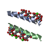
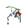
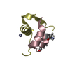
| Title | The crystal structures of the calcium-bound con-G and con-T(K7gamma) dimeric peptides demonstrate a novel metal-dependent helix-forming motif | ||||||
 Components Components | Conantokin-G | ||||||
 Keywords Keywords | METAL BINDING PROTEIN / conantoxin / con-G / NMDAR antagonist / Gla-containing | ||||||
| Function / homology | Conantokin, conserved site / Conantokin family signature. / host cell postsynaptic membrane / ion channel regulator activity / toxin activity / extracellular region / metal ion binding / Conantokin-G Function and homology information Function and homology information | ||||||
| Biological species |  Conus geographus (geography cone) Conus geographus (geography cone) | ||||||
| Method |  X-RAY DIFFRACTION / SYNCHROTRON / MOLECULAR REPLACEMENT / Resolution: 1.25 Å X-RAY DIFFRACTION / SYNCHROTRON / MOLECULAR REPLACEMENT / Resolution: 1.25 Å | ||||||
 Authors Authors | Cnudde, S.E. / Prorok, M. / Dai, Q. / Castellino, F.J. / Geiger, J.H. | ||||||
 Citation Citation | Journal: J.Am.Chem.Soc. / Year: 2007 Title: The crystal structures of the calcium-bound con-G and con-T[K7gamma] dimeric peptides demonstrate a metal-dependent helix-forming motif Authors: Cnudde, S.E. / Prorok, M. / Dai, Q. / Castellino, F.J. / Geiger, J.H. | ||||||
| History |
|
- Structure visualization
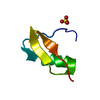
| Structure viewer | Molecule: MolmilJmol/JSmol |
|---|
- Downloads & links
-Download
| PDBx/mmCIF format | 2dpq.cif.gz | 23.8 KB | Display | PDBx/mmCIF format |
|---|---|---|---|---|
| PDB format | pdb2dpq.ent.gz | 15.7 KB | Display | PDB format |
| PDBx/mmJSON format | 2dpq.json.gz | Tree view | PDBx/mmJSON format | |
| Others |  Other downloads Other downloads |
-Validation report
| Arichive directory | https://data.pdbj.org/pub/pdb/validation_reports/dp/2dpqftp://data.pdbj.org/pub/pdb/validation_reports/dp/2dpq | HTTPS FTP |
|---|
-Related structure data

-Links
 PDBj
PDBj- Assembly
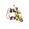
| Deposited unit | 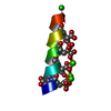
| ||||||||||||
|---|---|---|---|---|---|---|---|---|---|---|---|---|---|
| 1 | 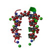
| ||||||||||||
| 2 | 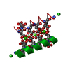
| ||||||||||||
| 3 | 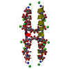
| ||||||||||||
| Unit cell |
| ||||||||||||
| Components on special symmetry positions |
| ||||||||||||
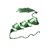
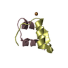
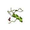
| Details | In the presence of calcium, con-G forms an antiparallel dimeric structure. |
-Components
| #1: Protein/peptide | Mass: 2265.196 Da / Num. of mol.: 1 / Source method: obtained synthetically Details: The con-G peptide was synthesized. The sequence of the peptide is naturally found in Conus geographus (geography cone) Source: (synth.) Conus geographus (geography cone) / References: UniProt: P07231 | ||
|---|---|---|---|
| #2: Chemical | ChemComp-CL /   Mass: 35.453 Da / Num. of mol.: 1 / Source method: obtained synthetically / Formula: Cl Mass: 35.453 Da / Num. of mol.: 1 / Source method: obtained synthetically / Formula: Cl | ||
| #3: Chemical |   Mass: 40.078 Da / Num. of mol.: 3 / Source method: obtained synthetically / Formula: Ca Mass: 40.078 Da / Num. of mol.: 3 / Source method: obtained synthetically / Formula: Ca#4: Water | ChemComp-HOH / |  Mass: 18.015 Da / Num. of mol.: 67 / Source method: isolated from a natural source / Formula: H2O Mass: 18.015 Da / Num. of mol.: 67 / Source method: isolated from a natural source / Formula: H2O |
-Experimental details
-Experiment
| Experiment | Method: X-RAY DIFFRACTION / Number of used crystals: 1 |
|---|
- Sample preparation
| Crystal | Density Matthews: 2.23 Å3/Da / Density % sol: 44.76 % |
|---|---|
| Crystal grow | Method: vapor diffusion, hanging drop / pH: 8 / Details: 35% dioxane, pH 8, VAPOR DIFFUSION, HANGING DROP |
-Data collection
| Diffraction | Mean temperature: 277 K |
|---|---|
| Diffraction source | Source: SYNCHROTRON / Site: APS  / Beamline: 32-ID / Wavelength: 1 Å / Beamline: 32-ID / Wavelength: 1 Å |
| Detector | Type: MAR CCD 165 mm / Detector: CCD / Date: May 12, 2003 |
| Radiation | Protocol: SINGLE WAVELENGTH / Monochromatic (M) / Laue (L): M / Scattering type: x-ray |
| Radiation wavelength | Wavelength: 1 Å / Relative weight: 1 |
| Reflection | Resolution: 1.2→50 Å / Num. obs: 12427 / % possible obs: 98 % / Observed criterion σ(F): 0 / Observed criterion σ(I): -3 / Rmerge(I) obs: 0.081 / Χ2: 1.036 / Net I/σ(I): 7.1 |
| Reflection shell | Resolution: 1.2→1.24 Å / Rmerge(I) obs: 0.923 / Num. unique all: 611 / Χ2: 0.353 / % possible all: 94.4 |
- Processing
| Software |
| ||||||||||||||||||||||||||||
|---|---|---|---|---|---|---|---|---|---|---|---|---|---|---|---|---|---|---|---|---|---|---|---|---|---|---|---|---|---|
| Refinement | Method to determine structure: MOLECULAR REPLACEMENT Starting model: 14-mer polyalanine helix Resolution: 1.25→8 Å / σ(F): 2 / Stereochemistry target values: Engh & Huber
| ||||||||||||||||||||||||||||
| Displacement parameters | Biso mean: 27.161 Å2 | ||||||||||||||||||||||||||||
| Refinement step | Cycle: LAST / Resolution: 1.25→8 Å
| ||||||||||||||||||||||||||||
| Refine LS restraints |
|
