 Movie
Movie Controller

[English] 日本語
 Yorodumi
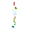
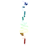
- PDB-2dp5: Structure of streptococcus pyogenes bacteriophage-associated hyal... -
+ Open data

- Basic information
| Entry | Database: PDB / ID: 2dp5 | ||||||
|---|---|---|---|---|---|---|---|
| Title | Structure of streptococcus pyogenes bacteriophage-associated hyaluronate lyase Hylp2 | ||||||
 Components Components | Hyaluronidase | ||||||
 Keywords Keywords | LYASE / HYALURONAN LYASE / PHAGE TAIL FIBRE / TRIPLE-STRANDED BETA-HELIX / HYALURONIDASE | ||||||
| Function / homology | Hyaluronidase, bacterial / Hyaluronidase protein (HylP) / Major tropism determinant, N-terminal domain / Major tropism determinant N-terminal domain / hyalurononglucosaminidase activity / capsule polysaccharide biosynthetic process / Hyaluronidase, phage associated Function and homology information Function and homology information | ||||||
| Biological species |  Streptococcus pyogenes (bacteria) Streptococcus pyogenes (bacteria) | ||||||
| Method |  X-RAY DIFFRACTION / MOLECULAR REPLACEMENT / Resolution: 3.55 Å X-RAY DIFFRACTION / MOLECULAR REPLACEMENT / Resolution: 3.55 Å | ||||||
 Authors Authors | Mishra, P. / Bhakuni, V. / Prem Kumar, R. / Singh, N. / Sharma, S. / Kaur, P. / Singh, T.P. | ||||||
 Citation Citation | Journal: To be Published Title: Structure of streptococcus pyogenes bacteriophage-associated hyaluronate lyase Hylp2 Authors: Mishra, P. / Bhakuni, V. / Prem Kumar, R. / Singh, N. / Sharma, S. / Kaur, P. / Singh, T.P. | ||||||
| History |
|
- Structure visualization
| Structure viewer | Molecule: MolmilJmol/JSmol |
|---|
- Downloads & links
-Download
| PDBx/mmCIF format | 2dp5.cif.gz | 74.3 KB | Display | PDBx/mmCIF format |
|---|---|---|---|---|
| PDB format | pdb2dp5.ent.gz | 56.2 KB | Display | PDB format |
| PDBx/mmJSON format | 2dp5.json.gz | Tree view | PDBx/mmJSON format | |
| Others |  Other downloads Other downloads |
-Validation report
| Arichive directory | https://data.pdbj.org/pub/pdb/validation_reports/dp/2dp5ftp://data.pdbj.org/pub/pdb/validation_reports/dp/2dp5 | HTTPS FTP |
|---|
-Related structure data
| Related structure data |  2c3fS S: Starting model for refinement |
|---|---|
| Similar structure data |
-Links
 PDBj
PDBj- Assembly
| Deposited unit | 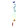
| ||||||||
|---|---|---|---|---|---|---|---|---|---|
| 1 | 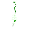
| ||||||||
| Unit cell |
|
-Components
| #1: Protein | Mass: 35834.176 Da / Num. of mol.: 1 Source method: isolated from a genetically manipulated source Source: (gene. exp.) Streptococcus pyogenes (bacteria) / Plasmid: PET 21d / Gene (production host): hyl P2 / Production host: |
|---|---|
| #2: Water | ChemComp-HOH /  Mass: 18.015 Da / Num. of mol.: 16 / Source method: isolated from a natural source / Formula: H2O Mass: 18.015 Da / Num. of mol.: 16 / Source method: isolated from a natural source / Formula: H2O |
-Experimental details
-Experiment
| Experiment | Method: X-RAY DIFFRACTION / Number of used crystals: 1 |
|---|
- Sample preparation
| Crystal | Density Matthews: 2.7 Å3/Da / Density % sol: 54.2 % |
|---|---|
| Crystal grow | Temperature: 298 K / Method: vapor diffusion, hanging drop / pH: 7.8 Details: Tris HCl, Sodium formate, pH 7.8, VAPOR DIFFUSION, HANGING DROP, temperature 298K |
-Data collection
| Diffraction | Mean temperature: 298 K |
|---|---|
| Diffraction source | Source: ROTATING ANODE / Type: RIGAKU RU300 / Wavelength: 1.5414 Å |
| Detector | Type: MARRESEARCH / Detector: IMAGE PLATE / Date: Mar 6, 2006 / Details: MIRROR |
| Radiation | Monochromator: GRAPHITE / Protocol: SINGLE WAVELENGTH / Monochromatic (M) / Laue (L): M / Scattering type: x-ray |
| Radiation wavelength | Wavelength: 1.5414 Å / Relative weight: 1 |
| Reflection | Resolution: 3.55→20 Å / Num. all: 5360 / Num. obs: 5360 / % possible obs: 98.1 % / Observed criterion σ(F): 0 / Observed criterion σ(I): 0 / Rsym value: 0.128 / Net I/σ(I): 4.7 |
| Reflection shell | Resolution: 3.55→3.65 Å / Mean I/σ(I) obs: 1.9 / Rsym value: 0.285 / % possible all: 97.8 |
- Processing
| Software |
| |||||||||||||||||||||||||||||||||||||||||||||||||||||||||||||||||||||||||||||||||||||||||||||||||||||||||||||||||||
|---|---|---|---|---|---|---|---|---|---|---|---|---|---|---|---|---|---|---|---|---|---|---|---|---|---|---|---|---|---|---|---|---|---|---|---|---|---|---|---|---|---|---|---|---|---|---|---|---|---|---|---|---|---|---|---|---|---|---|---|---|---|---|---|---|---|---|---|---|---|---|---|---|---|---|---|---|---|---|---|---|---|---|---|---|---|---|---|---|---|---|---|---|---|---|---|---|---|---|---|---|---|---|---|---|---|---|---|---|---|---|---|---|---|---|---|---|
| Refinement | Method to determine structure: MOLECULAR REPLACEMENT Starting model: 2C3F Resolution: 3.55→20 Å / Cor.coef. Fo:Fc: 0.91 / Cor.coef. Fo:Fc free: 0.885 / SU B: 45.134 / SU ML: 0.718 / Cross valid method: THROUGHOUT / σ(F): 0 / ESU R Free: 0.658 / Stereochemistry target values: MAXIMUM LIKELIHOOD / Details: HYDROGENS HAVE BEEN ADDED IN THE RIDING POSITIONS
| |||||||||||||||||||||||||||||||||||||||||||||||||||||||||||||||||||||||||||||||||||||||||||||||||||||||||||||||||||
| Solvent computation | Ion probe radii: 0.8 Å / Shrinkage radii: 0.8 Å / VDW probe radii: 1.4 Å / Solvent model: BABINET MODEL WITH MASK | |||||||||||||||||||||||||||||||||||||||||||||||||||||||||||||||||||||||||||||||||||||||||||||||||||||||||||||||||||
| Displacement parameters | Biso mean: 48.47 Å2
| |||||||||||||||||||||||||||||||||||||||||||||||||||||||||||||||||||||||||||||||||||||||||||||||||||||||||||||||||||
| Refinement step | Cycle: LAST / Resolution: 3.55→20 Å
| |||||||||||||||||||||||||||||||||||||||||||||||||||||||||||||||||||||||||||||||||||||||||||||||||||||||||||||||||||
| Refine LS restraints |
| |||||||||||||||||||||||||||||||||||||||||||||||||||||||||||||||||||||||||||||||||||||||||||||||||||||||||||||||||||
| LS refinement shell | Resolution: 3.55→3.639 Å / Total num. of bins used: 20 /
|
