 Movie
Movie Controller

[English] 日本語
 Yorodumi
- PDB-2dh1: Crystal structure of peanut lectin lactose-azobenzene-4,4'-dicarb... -
+ Open data

- Basic information
| Entry | Database: PDB / ID: 2dh1 | ||||||
|---|---|---|---|---|---|---|---|
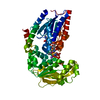
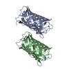
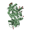
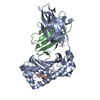
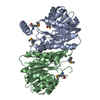
| Title | Crystal structure of peanut lectin lactose-azobenzene-4,4'-dicarboxylic acid-lactose complex | ||||||
 Components Components | Galactose-binding lectin | ||||||
 Keywords Keywords | SUGAR BINDING PROTEIN / LEGUME LECTIN / AGGLUTININ / CROSSLINK / OPEN QUATERNARY STRUCTURE / CARBOHYDRATE SPECIFICITY / MULTIVALENCY | ||||||
| Function / homology |  Function and homology information Function and homology information | ||||||
| Biological species |  | ||||||
| Method |  X-RAY DIFFRACTION / MOLECULAR REPLACEMENT / Resolution: 7.65 Å X-RAY DIFFRACTION / MOLECULAR REPLACEMENT / Resolution: 7.65 Å | ||||||
 Authors Authors | Natchiar, S.K. / Srinivas, O. / Nivedita, M. / Sagarika, D. / Jayaraman, N. / Surolia, A. / Vijayan, M. | ||||||
 Citation Citation | Journal: Curr.Sci. / Year: 2006 Title: Multivalency in lectins - A crystallographic, modelling and light-scattering study involving peanut lectin and a bivalent ligand Authors: Natchiar, S.K. / Srinivas, O. / Nivedita, M. / Sagarika, D. / Jayaraman, N. / Surolia, A. / Vijayan, M. #1: Journal: Proc.Natl.Acad.Sci.Usa / Year: 1994 Title: Crystal structure of peanut lectin, a protein with an unusual quaternary structure Authors: Banerjee, R. / Mande, S.C. / Ganesh, V. / Das, K. / Dhanaraj, V. / Mahanta, S.K. / Suguna, K. / Surolia, A. / Vijayan, M. #2: Journal: J.Mol.Biol. / Year: 1996Title: Conformation, protein-carbohydrate interactions and a novel subunit association in the refined structure of peanut lectin-lactose complex Authors: Banerjee, R. / Das, K. / Ravishankar, R. / Suguna, K. / Surolia, A. / Vijayan, M. #3: Journal: Curr.Sci. / Year: 1997Title: The Specificity of Peanut Agglutinin for Thomsen-Friedenreich Antigen is Mediated by Water-Bridges Authors: Ravishankar, R. / Ravindran, M. / Suguna, K. / Surolia, A. / Vijayan, M. #4: Journal: ACTA CRYSTALLOGR.,SECT.D / Year: 2004Title: Structural plasticity of peanut lectin: an X-ray analysis involving variation in pH, ligand binding and crystal structure Authors: Natchiar, S.K. / Jeyaprakash, A.A. / Ramya, T.N. / Thomas, C.J. / Suguna, K. / Surolia, A. / Vijayan, M. #5: Journal: J.Am.Chem.Soc. / Year: 2002 Title: Photoswitchable multivalent sugar ligands: synthesis, isomerization, and lectin binding studies of azobenzene-glycopyranoside derivatives Authors: Srinivas, O. / Mitra, N. / Surolia, A. / Jayaraman, N. | ||||||
| History |
|
- Structure visualization
| Structure viewer | Molecule: MolmilJmol/JSmol |
|---|
- Downloads & links
-Download
| PDBx/mmCIF format | 2dh1.cif.gz | 40.3 KB | Display | PDBx/mmCIF format |
|---|---|---|---|---|
| PDB format | pdb2dh1.ent.gz | 25.6 KB | Display | PDB format |
| PDBx/mmJSON format | 2dh1.json.gz | Tree view | PDBx/mmJSON format | |
| Others |  Other downloads Other downloads |
-Validation report
| Arichive directory | https://data.pdbj.org/pub/pdb/validation_reports/dh/2dh1ftp://data.pdbj.org/pub/pdb/validation_reports/dh/2dh1 | HTTPS FTP |
|---|
-Related structure data
| Related structure data | 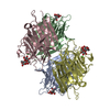 2pelS S: Starting model for refinement |
|---|---|
| Similar structure data |


-Links
 PDBj
PDBj

- Assembly
| Deposited unit | 
| ||||||||
|---|---|---|---|---|---|---|---|---|---|
| 1 | 
| ||||||||
| 2 | 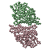
| ||||||||
| Unit cell |
| ||||||||
| Details | Biological molecule is a tetramer. It can be generated from the dimer in the asymmetric by symmetry |
-Components
| #1: Protein | Mass: 25208.955 Da / Num. of mol.: 4 / Source method: isolated from a natural source / Source: (natural) |
|---|
-Experimental details
-Experiment
| Experiment | Method: X-RAY DIFFRACTION / Number of used crystals: 1 |
|---|
- Sample preparation
| Crystal | Density Matthews: 4.6 Å3/Da / Density % sol: 73.2 % |
|---|---|
| Crystal grow | Temperature: 293 K / Method: vapor diffusion, hanging drop / pH: 7 Details: 12% PEG 8000, 0.05M sodium phosphate, 0.2M sodium cholride, 0.02% sodium azide, pH 7.0, VAPOR DIFFUSION, HANGING DROP, temperature 293.0K |
-Data collection
| Diffraction | Mean temperature: 293 K |
|---|---|
| Diffraction source | Source: ROTATING ANODE / Type: RIGAKU RU200 / Wavelength: 1.5418 / Wavelength: 1.5418 Å |
| Detector | Type: MARRESEARCH / Detector: IMAGE PLATE / Date: Oct 3, 2003 / Details: OSMIC MIRROR |
| Radiation | Protocol: SINGLE WAVELENGTH / Monochromatic (M) / Laue (L): M / Scattering type: x-ray |
| Radiation wavelength | Wavelength: 1.5418 Å / Relative weight: 1 |
| Reflection | Resolution: 7.65→20 Å / Num. obs: 2273 / % possible obs: 99.8 % / Redundancy: 8.1 % / Rmerge(I) obs: 0.1 / Rsym value: 0.1 |
| Reflection shell | Resolution: 7.65→7.8 Å / Redundancy: 8.1 % / Rmerge(I) obs: 0.49 / Mean I/σ(I) obs: 4.6 / Num. unique all: 219 / Rsym value: 0.524 / % possible all: 100 |
- Processing
| Software |
| ||||||||||||||||||||
|---|---|---|---|---|---|---|---|---|---|---|---|---|---|---|---|---|---|---|---|---|---|
| Refinement | Method to determine structure: MOLECULAR REPLACEMENT Starting model: PDB ENTRY 2PEL Resolution: 7.65→20 Å / Cor.coef. Fo:Fc: 0.812 / Cor.coef. Fo:Fc free: 0.836 / SU B: 897.888 / SU ML: 6.067 / ESU R: 4.956 / ESU R Free: 5.272 / Stereochemistry target values: MAXIMUM LIKELIHOOD Details: RIGID BODY REFINEMENT. THE COORDINATES FOR ONLY THE ALPHA CARBONS ARE PRESENT IN THE STRUCTURE. THE NUMBER OF MISSING ATOMS WAS SO MUCH THAT REMARK 470 FOR THE MISSING ATOMS LIST WERE REMOVED.
| ||||||||||||||||||||
| Solvent computation | Ion probe radii: 0.8 Å / Shrinkage radii: 0.8 Å / VDW probe radii: 1.2 Å / Solvent model: BABINET MODEL WITH MASK | ||||||||||||||||||||
| Refinement step | Cycle: LAST / Resolution: 7.65→20 Å
| ||||||||||||||||||||
| LS refinement shell | Resolution: 7.65→7.91 Å / Total num. of bins used: 20
|
